Anatomy
Origin:
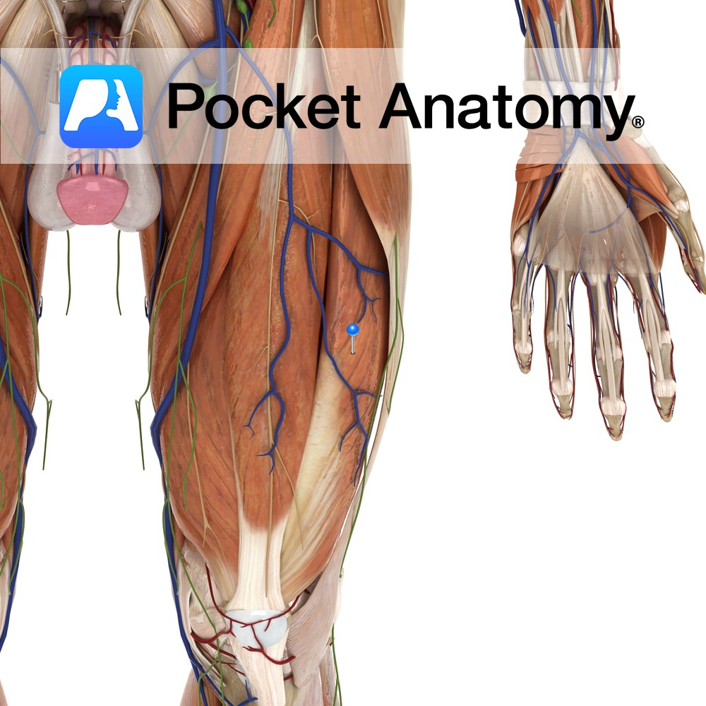
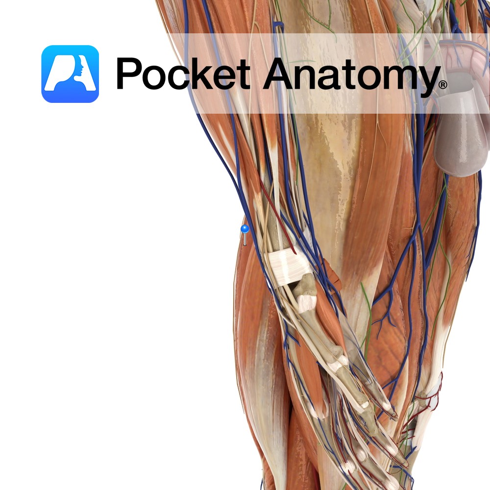
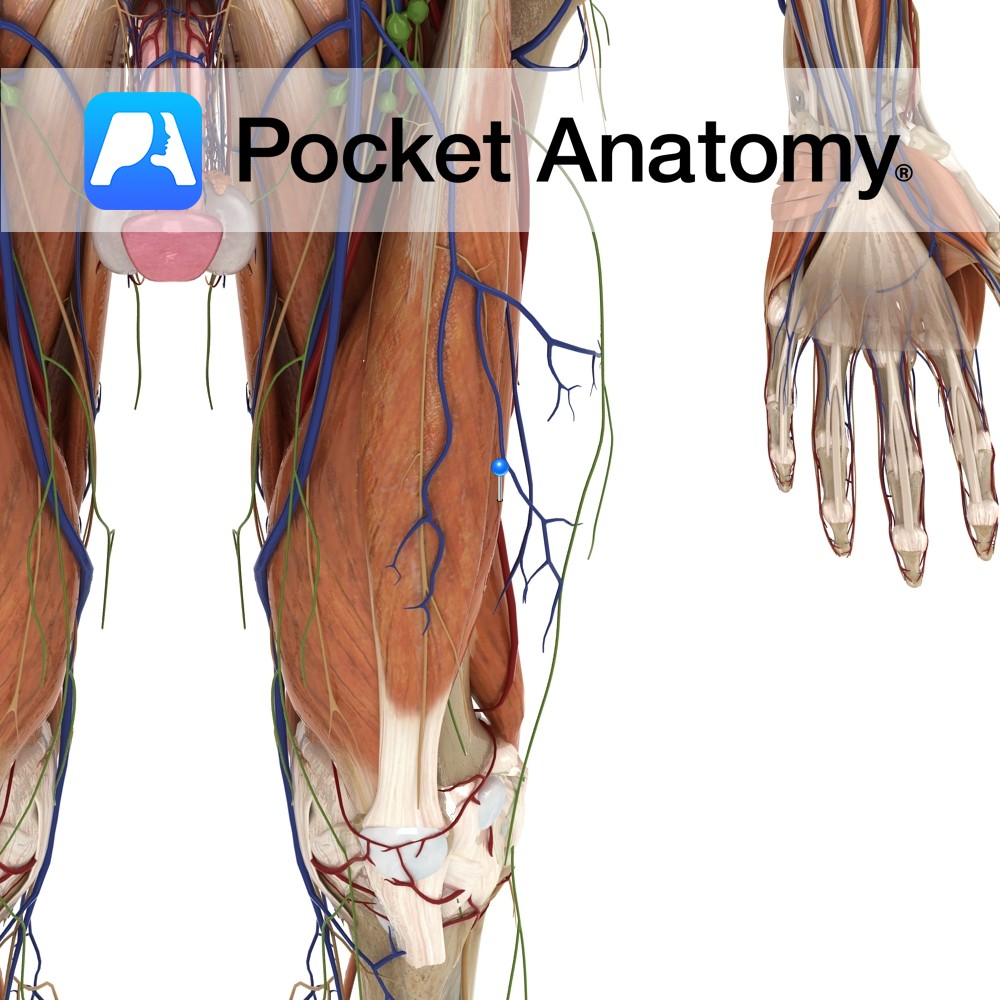
Greater trochanter and lateral lip of linea aspera of femur.
Insertion:
Base of the patella via the quadriceps femoris tendon. The quadriceps femoris tendon is functionally continuous with the patellar ligament which runs from the apex of the patella to the tibial tuberosity.
Key Relations:
-One of the five muscles of the anterior compartment of the thigh.
-Largest of the vastus muscles.
Note:
The quadriceps femoris is a large muscle group containing the 3 vastus muscles and the rectus femoris muscle of the anterior thigh. The quadriceps femoris tendon represents their shared insertion onto the patella.
Functions
Together with the other muscles that make up quadriceos femoris extends the leg at the knee joint.
Supply
Nerve Supply:
Femoral nerve (L2, L3, L4).
Blood Supply:
-Lateral circumflex femoral artery
-Deep femoral artery.
Clinical
The posterior surface of the patella is covered by layer of smooth articular cartilage. This allows the patella to glide smoothly over the articular groove of the femur as the knee flexes. While cartilage degeneration occurs with age (for example the degeneration seen with osteoarthritis), excessive or uneven pressure on the surface can also cause a degree of damage to the surface, causing pain. This particular syndrome is referred to as patellofemoral pain syndrome or anterior knee pain syndrome.
One cause of uneven pressure on the patella is an imbalance of the quadriceps femoris muscles of the anterior thigh, in particular vastus medialis and vastus lateralis. In teenagers the vastus lateralis has a tendency to be more powerful than vastus medialis. This causes an unbalanced force on the patella which tends to be pulled laterally when the leg is extended. Excessive pressure is put on the lateral facet of the patella where it can cause breakdown of the cartilage and knee pain especially when making knee movements. In the majority of patients this pain comes and goes, but usually resolves completely when they finish growing.
Interested in taking our award-winning Pocket Anatomy app for a test drive?


