Anatomy
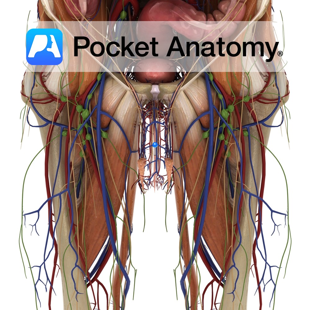
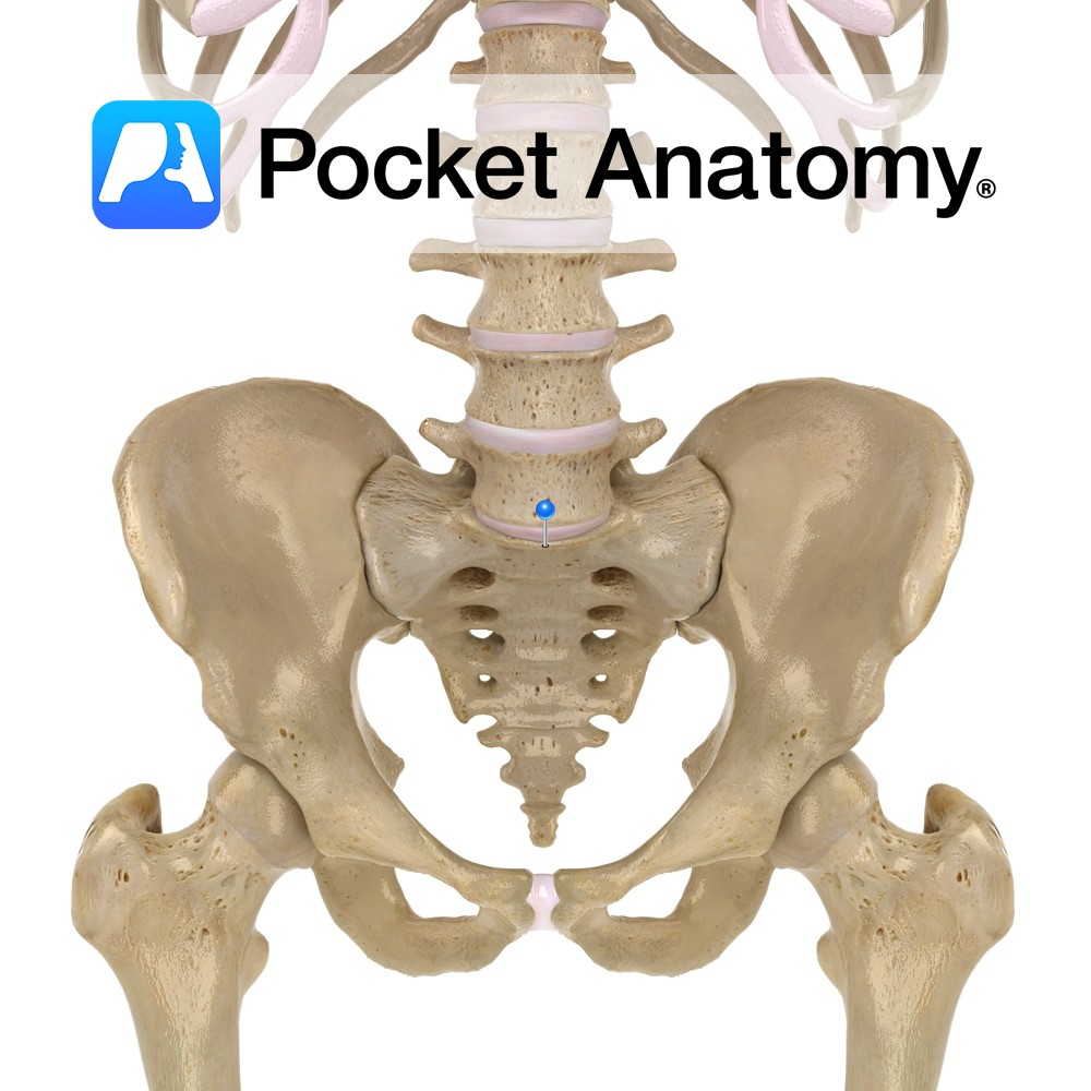
20cm muscular tube/conduit (female 4cm), extending down from the internal urethral orifice (apex of trigone in floor/neck of bladder, behind pubic symphysis) to external urethral orifice (meatus, at tip of glans) at end of penis.
There are two functional parts:
1. posterior/sphincter-active (made up of 3 sections – pre-prostatic/intramural in bladder, prostatic, membranous in external urethral sphincter)
2. anterior/spongy (coursing distally/down through (3) bulbar, penile, navicular/glans parts of corpus spongiosum).
Lumen about 0.5cm, expanded at each end (bulb and glans) to 1-1.5cm.
Physiology
Lumen flattened side-to-side other than in glans where vertical (hence “tight” spiral urine stream), smooth muscle collar at level of bladder neck (extension of detrusor muscle).
Clinical
Urethritis, when infective, is usually caused either by bacteria associated with urinary tract infection (UTI), or those associated with sexually transmitted disease (STD) such as gonorrhea, chlamydia. At risk men; 20-35, multiple partners, unprotected/anal intercourse. Symptoms include pain/burning, frequency, discharge. Can be chronic and non-infective.
Interested in taking our award-winning Pocket Anatomy app for a test drive?


.jpg)
