Anatomy
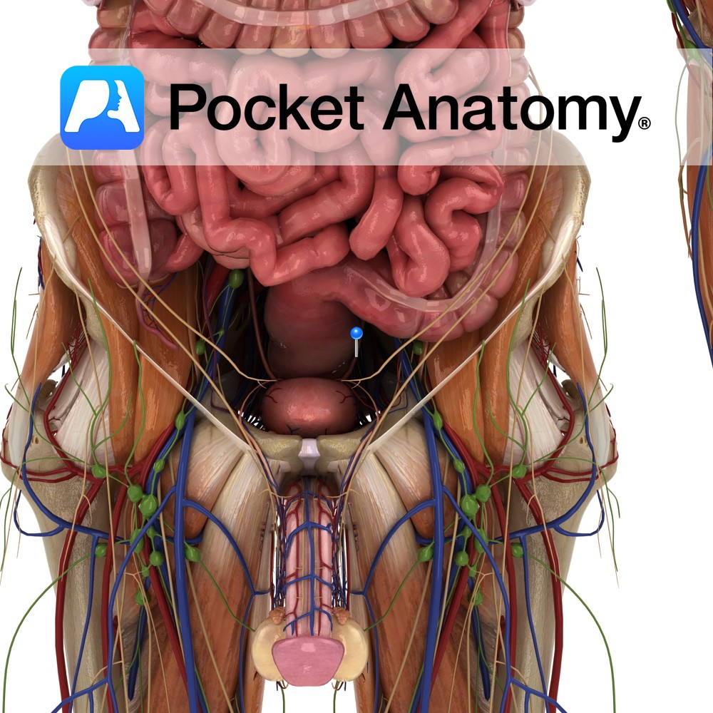
Paired autonomically innervated muscular tubes (25cms long, 0.5cm diameter) connecting kidney (medial aspect, at and continuous with renal pelvis, at level 2nd Lumbar vertebra) to bladder (upper aspect posterior/base, at level of pelvic tubercle), through which urine is propelled.
Ureter courses down and medially, behind the peritoneum, in front of psoas, crossing pelvic brim in front of bifurcation of common iliac artery, medial to sacriliac joint, at which point ureters 5 cms apart, then diverge laterally almost to ischial spine, then back in medially to base of bladder (at level pubic tubercle). Behind it; psoas, genitofemoral nerve, common iliac, SI joint. In front of it; gonadal and colic vessels. Multiple arterial supply, to its medial aspect in abdomen, to its lateral aspect in pelvis. Urine propelled by peristalsis, controlled by intrinsic pacemaker.
Clinical
Ureteric stones (urolithiasis) common (12% men by 70, 6% women), 90% detectable on plain X-ray, 85% eventually passed through urination/micturition, associated sudden pain severe and colicy (referred; from highest part to flank, lower to front lower abdomen, then groin, scrotum/labia, upper/inner thigh), 3 commonest sites of blockage – ureteropelvic junction, ureterovesical valve, point at which crosses in front of common iliac (or its bifurcation).
Ureteric damage (ligation or transection) a constant concern in general, gynaecologic, vascular surgery. Blockage also possible with constipation, gravid uterus, tumour.
Interested in taking our award-winning Pocket Anatomy app for a test drive?


