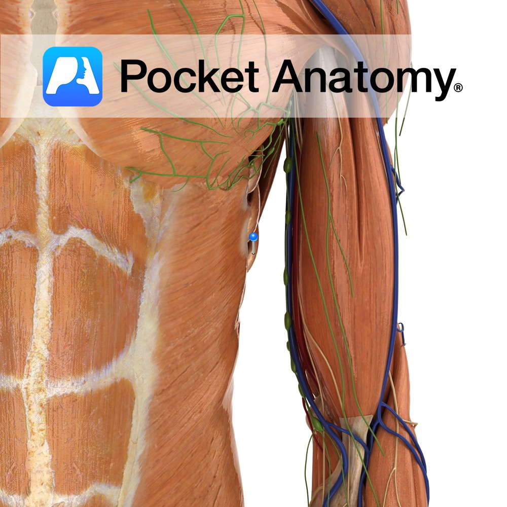
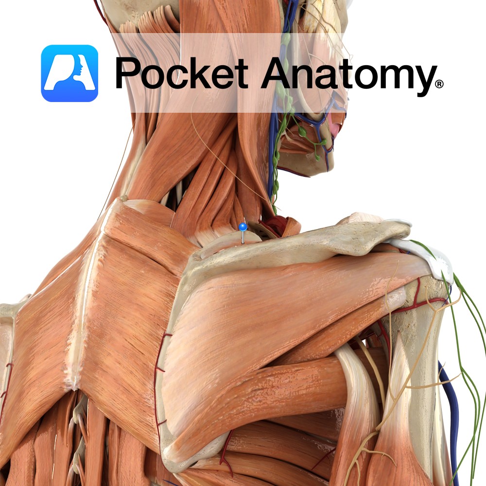
Anatomy
Origin:
External surface of lateral surface of upper eight ribs.
Insertion:
Medial border of scapula.
Key Relations:
The long thoracic nerve travels inferiorly on the surface of the muscle.
Functions
-Protracts the scapula laterally rotates the scapula e.g. as in the forward motion of throwing a punch.
-Stabilises and holds the medial border of the scapula against the thoracic wall.
-Laterally rotates the scapula.
Supply
Nerve Supply:
Innervated by the long thoracic nerve (C5, C6, C7).
Blood Supply:
-Superior thoracic artery
–Lateral thoracic artery
-Thoracodorsal artery.
Clinical
Paralysis or weakness of the muscle causes the medial border of the scapula to project backwards known as ‘winging’ of the scapula.
The serratus anterior can be tested by asking the patient to stand just less than their arms’ length away from a wall and to get them to push against the wall. The digitations of the muscle may be seen and felt against the lateral thoracic wall. This may be more visible in young or athletic patients. Another test for this muscle is to ask the patient to do a ‘press-up’ and inspect the same region.
Interested in taking our award-winning Pocket Anatomy app for a test drive?


