Anatomy
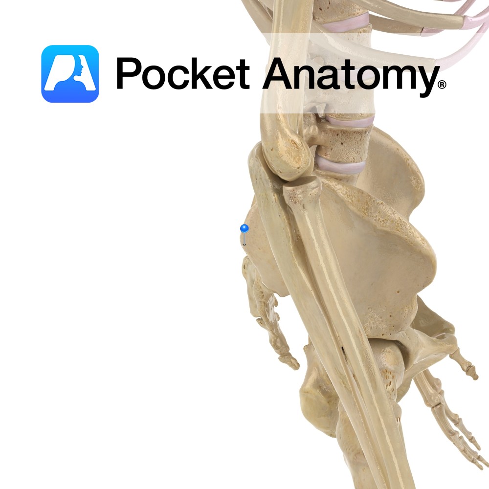
Origin:
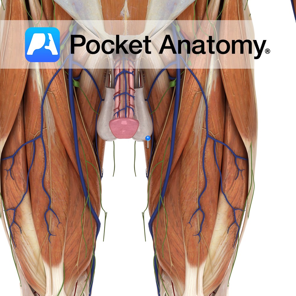
Body and inferior ramus of pubis.
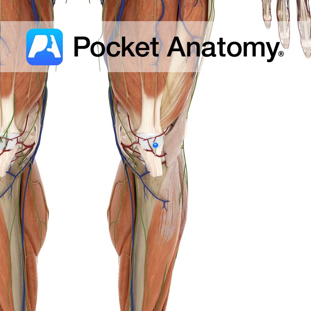
Insertion:
Superomedial surface of tibia.
Key Relations:
-One of the six muscles of the medial compartment of the thigh.
-At its insertion to the tibia, the tendon of gracilis is located between the tendons of sartorius and semitendinosus. This 3 pronged arrangement is known as pes anserinus (the latin for goosefoot).
Functions
-Adducts thigh at the hip joint.
-Flexes and medially rotates the leg at the knee joint.
Supply
Nerve Supply:
Obturator nerve (L2, L3).
Blood Supply:
–Obturator artery
-Medial circumflex femoral artery.
Clinical
One surgical option for the treatment of faecal incontinence involves transposition of one of the gracilis muscles in a loop around the external surface of the anal canal. An electrical pacemaker keeps the muscle contracted therefore maintaining continence. The pacemaker can be switched off using an external magnet thus allowing defaecation. This procedure has proven to be quite successful in restoring continence and improving the quality of life of patients with faecal incontinence.
The adductor muscles of the thigh are tested as a functional group. The patient is requested to lie with their knees extended on the opposite side of that to be tested. The physician will abduct the leg on the side being tested, and then ask the patient to adduct the leg against resistance.
Interested in taking our award-winning Pocket Anatomy app for a test drive?


