Anatomy
Origin:
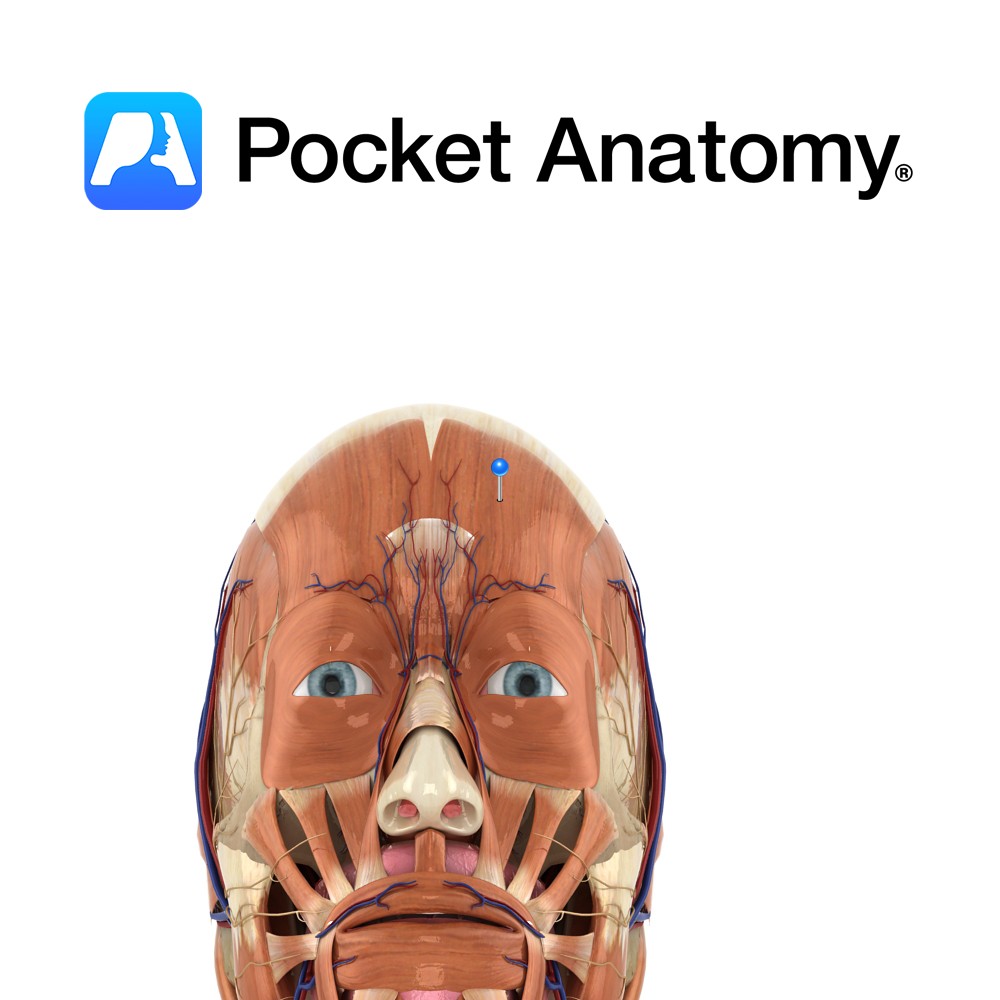
Frontalis has no bony origin. Its fibres are continuous with those of the muscles procerus and corrgator supercilii.
Insertion:
Fibres join the galea aponeurotica (epicranial aponeurosis) below the coronal suture.
Key Relations:
Many consider frontalis and occipitalis as one muscle (called occipitofrontalis) with an aponeurotic tendon between two bellies- a frontal belly and an occipital belly.
Functions
Elevates eyebrows and wrinkles forehead.
e.g. as in looking surprised..
Supply
Nerve Supply:
Temporal branch of the facial nerve (CN 7).
Blood Supply:
Opthalmic artery.
Clinical
Frontalis receives bilateral innervation. This means that its actions are relatively spared in an upper motor neuron (e.g. stroke) lesion and the patient will still be able to wrinkle their whole forehead. All strength is lost in the muscle ipsilaterally in a lower motor neuron lesion (e.g. Bell’s palsy) the other half of the muscle only will have the ability to wrinkle its half of the forehead. This differentiation is vital in differentiating at which level a lesion exists in patients with facial weakness.
Interested in taking our award-winning Pocket Anatomy app for a test drive?


