Anatomy
Origin:
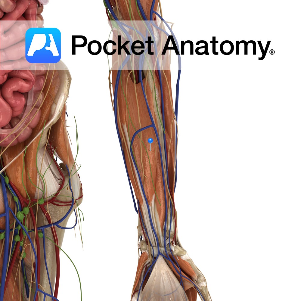
Upper three quarters of medial and anterior aspects of ulna and from anterior aspect of interosseous membrane.
Insertion:
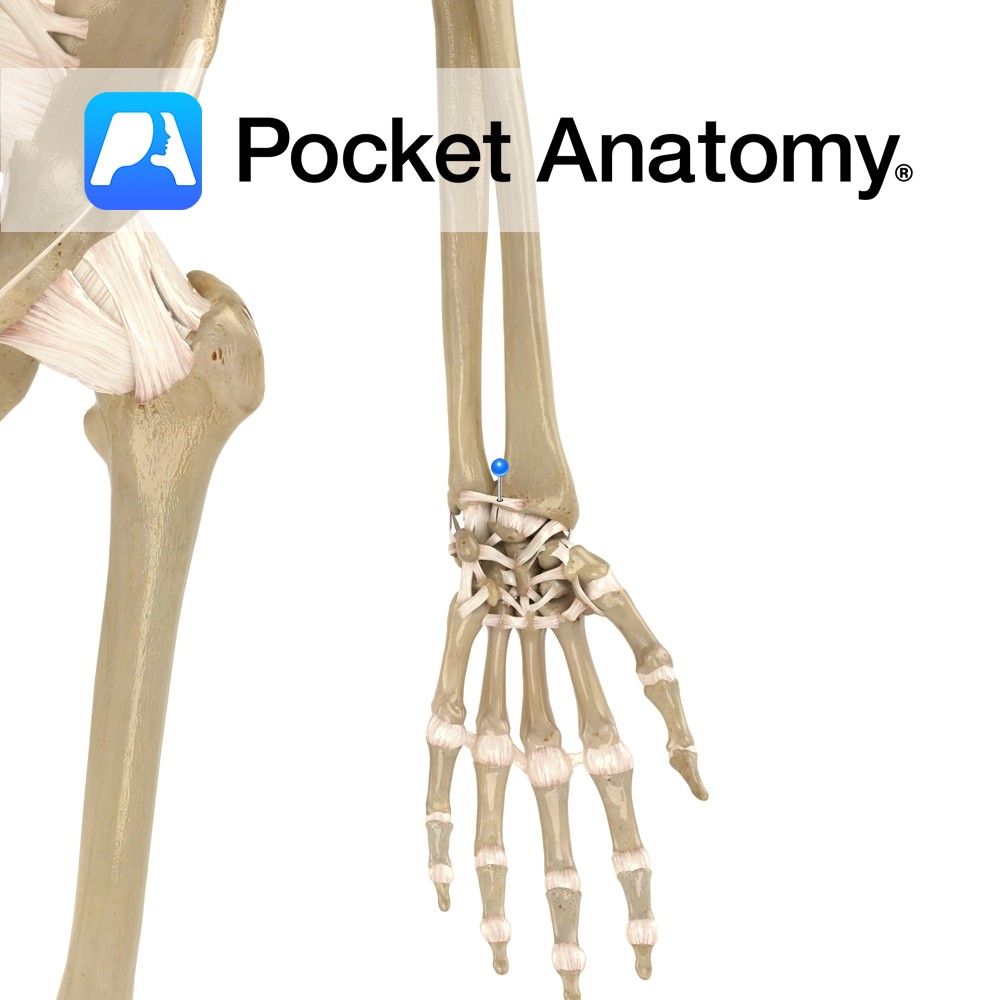
Four tendons attaching to palmar aspects of bases of the distal phalanges of each of the four fingers.
Key Relations:
-The lumbrical muscles arise in the palm from the tendons of flexor digitorum profundus.
-The four tendons of the muscle pass through the carpal tunnel as they enter the hand.
-To reach the distal phalanx the tendons of flexor digitorum profundus pass beneath the fibrous tunnels formed by flexor digitorum superficialis.
-One of the three muscles in the deep anterior compartment of the forearm.
Functions
-The sole flexor of the distal interphalangeal joints of the four fingers.
-Flexes the proximal interphalangeal joints and metacarpophalangeal joints of the fingers (along with flexor digitorum superficialis).
-Helps flex the hand at the wrist
e.g. when making a fist.
Supply
Nerve Supply:
Medial half: (little finger, ring finger): ulnar nerve (C8,T1).
Lateral half: (index finger, middle finger): Anterior interosseous nerve (C8,T1) – a branch of the median nerve.
Blood Supply:
Anterior interosseous artery.
Clinical
Tearing away of the tendon from its insertion may occur with forced extension of the fingers from an initial gripping or flexed position. As the flexor digitorum profundus is the only muscle involved in flexion of the distal interphalangeal joints this injury can only be picked up by checking a patient’s ability to flex these joints.
Interested in taking our award-winning Pocket Anatomy app for a test drive?


