Anatomy
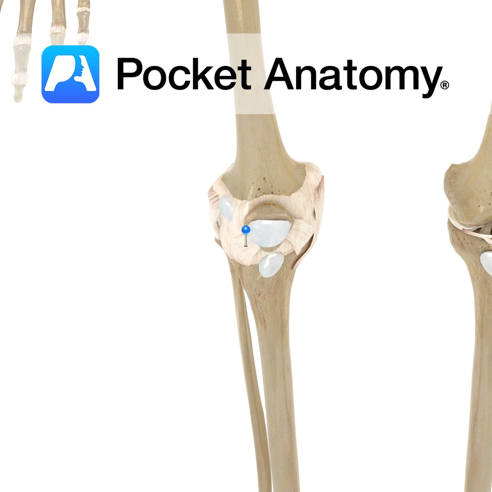
Origin:
Inferior aspect of lateral condyle of tibia, upper three-fourths of the anterior surface of the interosseous membrane and the anterior intermuscular septum.
Insertion:
The extensor digitorum longus tendon divides into four slips deep to the inferior extensor retinaculum. The four slips insert onto the dorsal aspects of the middle and distal phalanges of the lateral four digits via the dorsal digital expansions.
Key Relations:
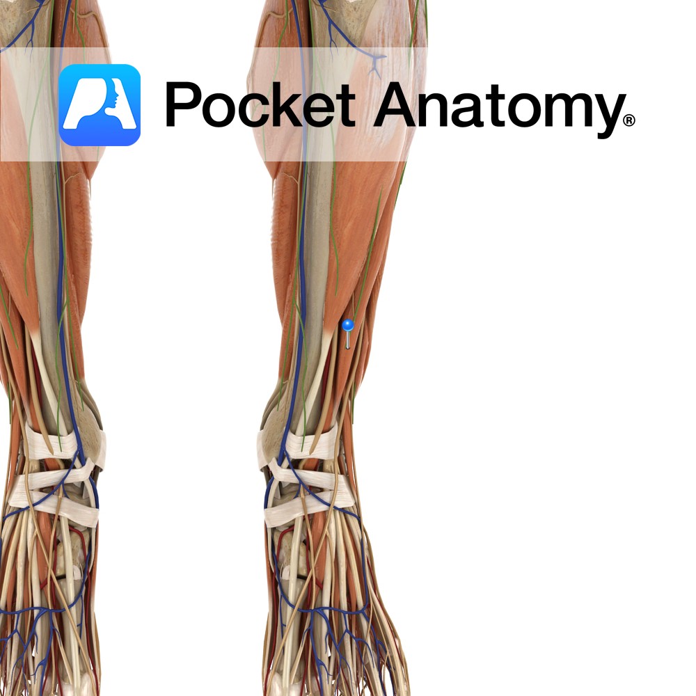
-One of the four muscles of the anterior compartment of the leg.
-The extensor digitorum longus tendon crosses anterior to the ankle joint lateral to the extensor hallucis longus tendon.
Functions
-Extends the lateral four digits at the interphalangeal and metatarsophalangeal joints e.g. making sure the toes clear the steps when walking up a stairs.
-Dorsiflexes the ankle joint.
-Also tautens the plantar aponeurosis.
Supply
Nerve Supply:
Deep fibular (peroneal) nerve (L5, S1).
Blood Supply:
Anterior tibial artery.
Clinical
Extensor tendinitis is inflammation of the extensor tendons of the toes such as tibialis anterior, extensor hallucis longus, extensor hallucis brevis, extensor digitorum longus and extensor digitorum brevis. Although inflammation of the extensor digitorum muscles is rare. The patient may present with pain and swelling on the dorsum of the foot, which may be worsened by activity e.g running or by passive stretching of the tendons.
Extensor tendinitis may be caused by overuse, hill running as the extensor muscles have to work harder, tight-fitted shoes or if shoe laces are tied too tightly. Treatment includes rest, ice, NSAIDs, better fit shoes, stretching the calf muscles and in severe cases surgery.
Interested in taking our award-winning Pocket Anatomy app for a test drive?
![]()
.jpg)