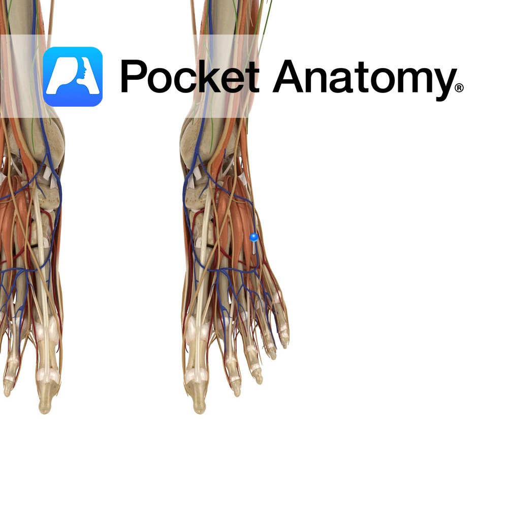
Anatomy
Origin:
Anterolateral part of the superior surface of calcaneus and inferior extensor retinaculum.
Insertion:
Lateral aspect of the extensor digitorum longus tendons of 2nd, 3rd and 4th toes.
Key relations:
Runs anteromedially across the foot and can be palpated inferomedially to the lateral malleolus on the dorsum of the foot.
Functions
Extends the 2nd, 3rd and 4th toes (assists extensor digitorum longus) at the metatarsophalangeal joints.
Supply
Nerve supply:
Deep fibular (peroneal) nerve (L5,S1)
Blood supply:
-Peroneal (fibular) artery
-Dorsalis pedis artery (continuation of anterior tibial artery).
Interested in taking our award-winning Pocket Anatomy app for a test drive?


