Anatomy
Course
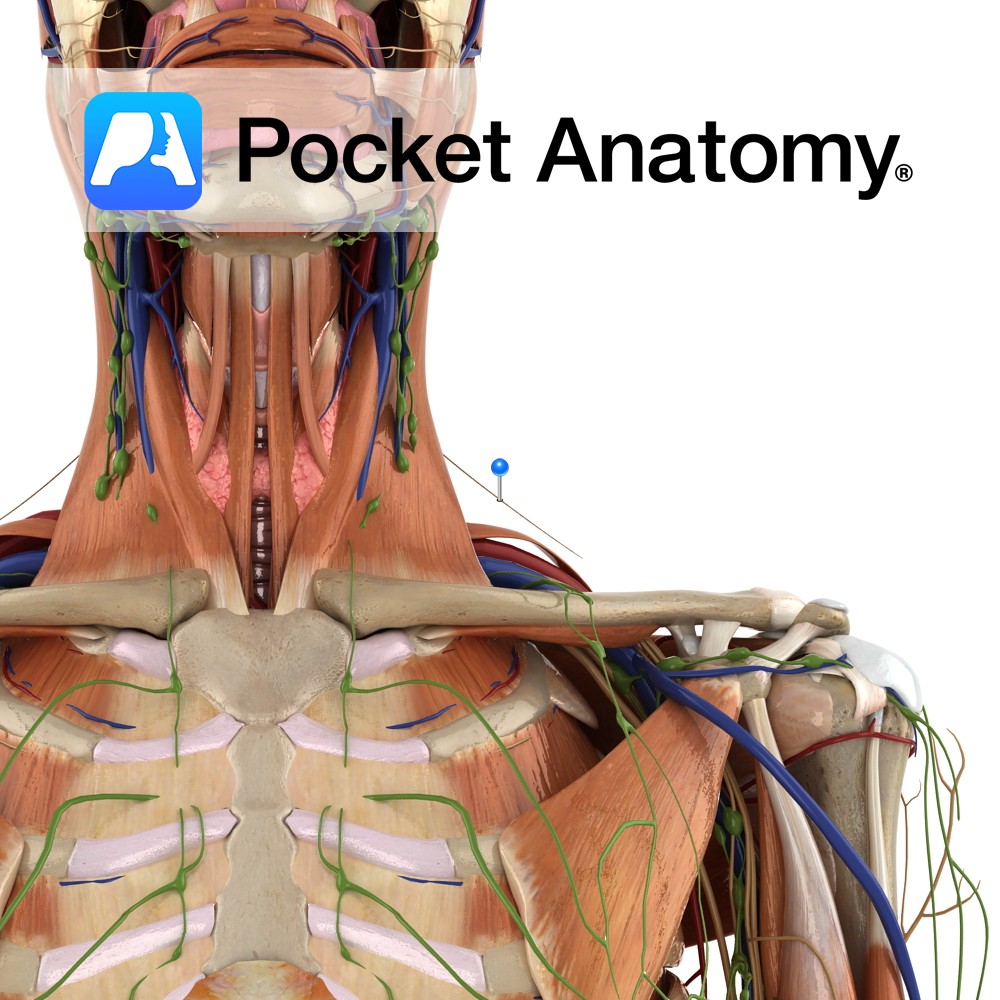
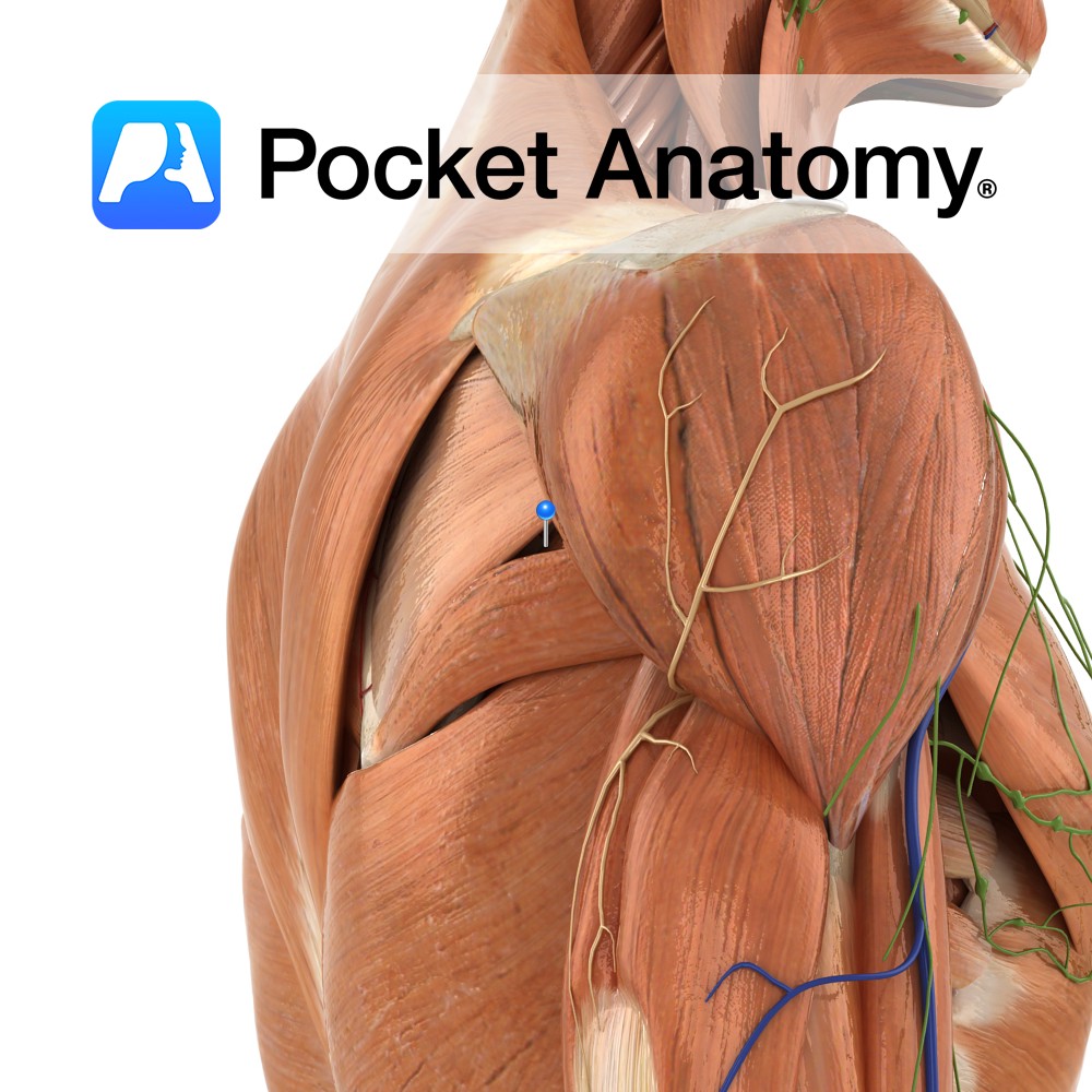
Also known as the eleventh cranial nerve, its spinal root enters the cranium via the foramen magnum to join the cranial root, and it then exits via the jugular foramen along with the glossopharyngeal and vagus nerves. Once out of the cranium, the cranial portion of the nerve dissociates and joins the vagus nerve. During its descent down the neck, it crosses the internal jugular vein. It eventually pierces the sternocleidomastoid muscle but continues to travel until it reaches the trapezius.
Supply
Motor innervation of the trapezius muscles and the sternocleidomastoid muscle.
Clinical
The distal portion of the nerve is most prone to injury due to its exposed nature. Damage to the nerve is usually seen in the clinic as one-sided shoulder weakness, identified by having the patient shrug their shoulders against resistance.
Interested in taking our award-winning Pocket Anatomy app for a test drive?


