PocketAnatomy® is a registered brand name owned by © eMedia Interactive Ltd, 2009-2022.
iPhone, iPad, iPad Pro and Mac are trademarks of Apple Inc., registered in the U.S. and other countries. App Store is a service mark of Apple Inc.
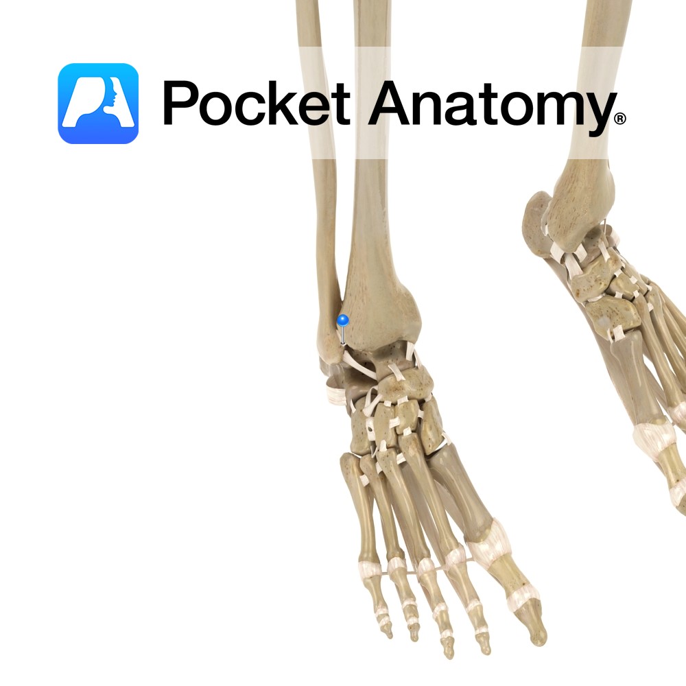
Anatomy Part of the distal tibiofibular joint. It attaches from the lateral surface of the anterior distal tibia, and travels obliquely downwards to attach to the lateral malleolus. Functions Holds the distal end of the tibiofibular joint together tightly. It is key for the skeletal framework and articulation of the foot at the ankle. Interested
- Published in Pocket Anatomy Pins
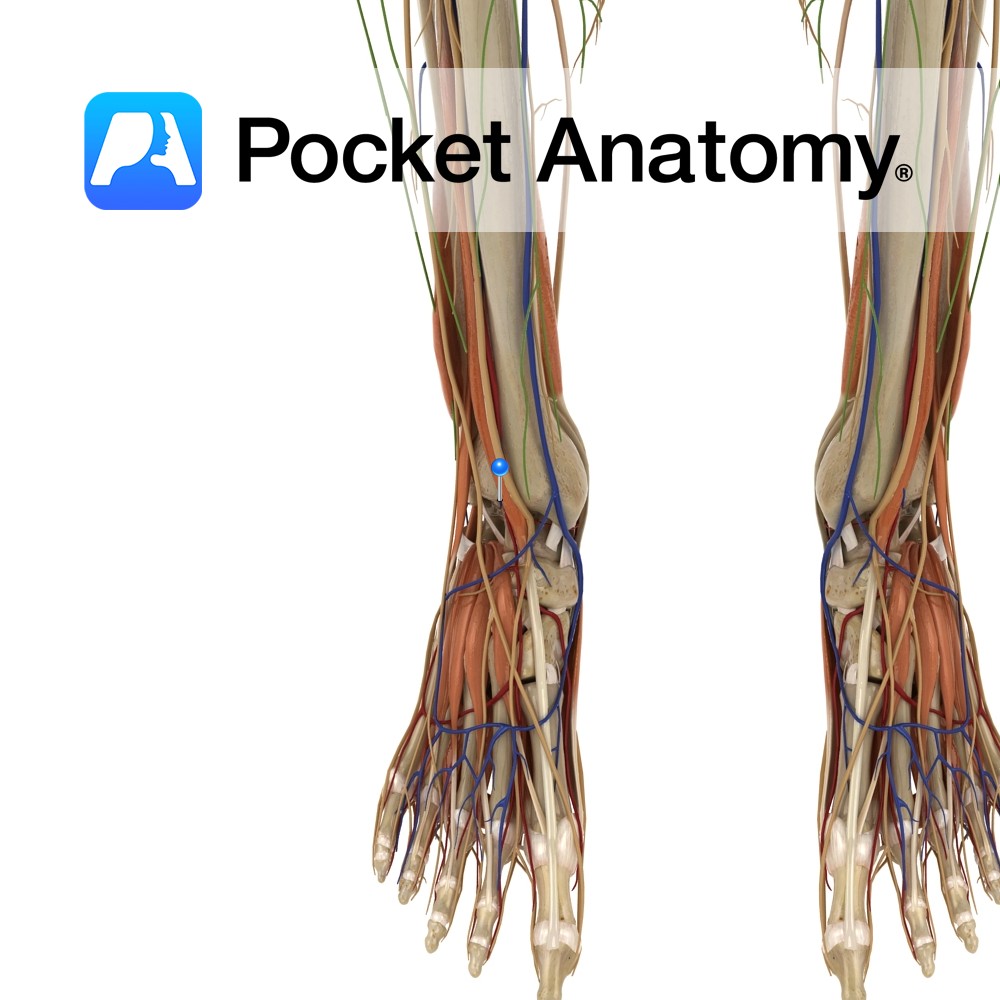
Anatomy Course Follows its counterpart artery until it drains into the popliteal vein. Drain Drains the anterior compartment of the lower limb. Interested in taking our award-winning Pocket Anatomy app for a test drive?
- Published in Pocket Anatomy Pins
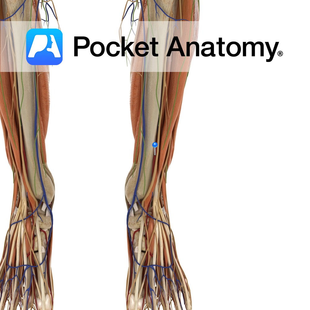
Anatomy Course Continuation of the popliteal artery when it passes through the interosseous membrane to cross into the anterior leg. It then descends on the anterior side of the interosseous membrane until it reaches the distal end of the tibia and ankle. It then continues into the foot as the dorsalis pedis artery. Supply Supplies
- Published in Pocket Anatomy Pins
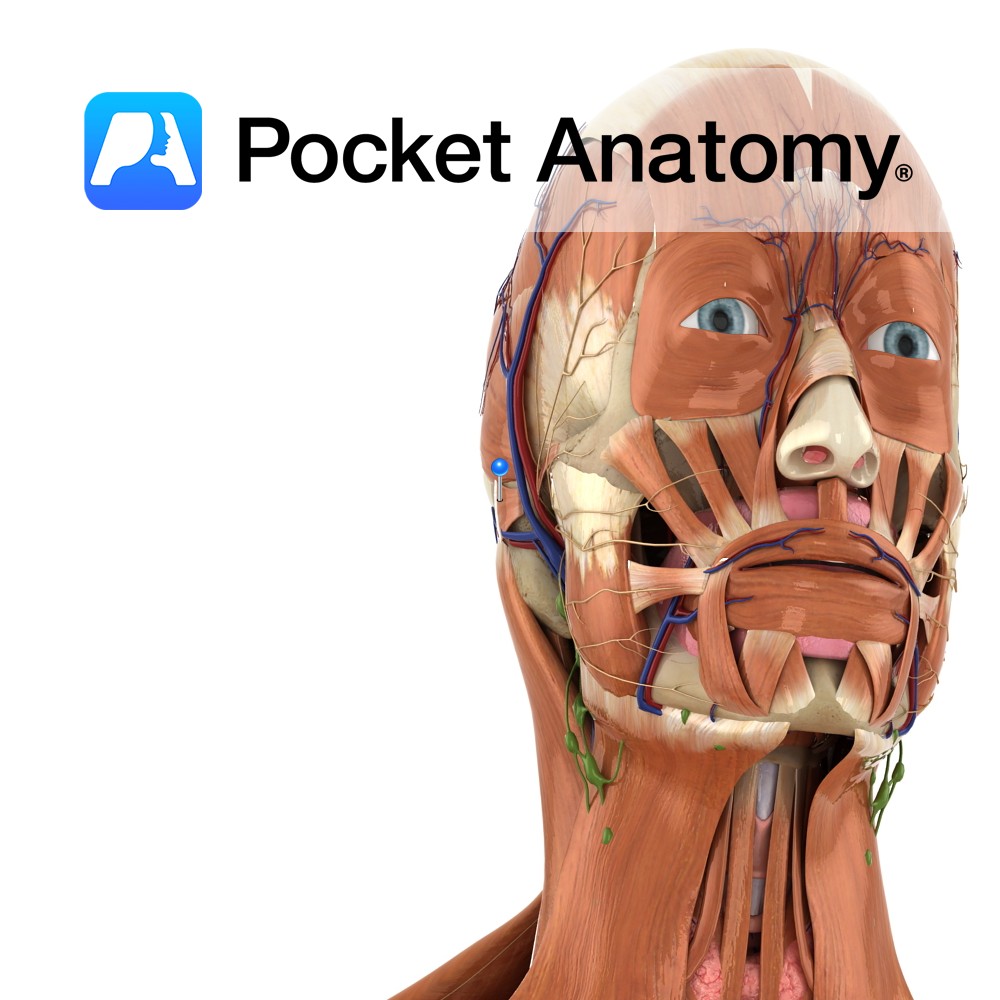
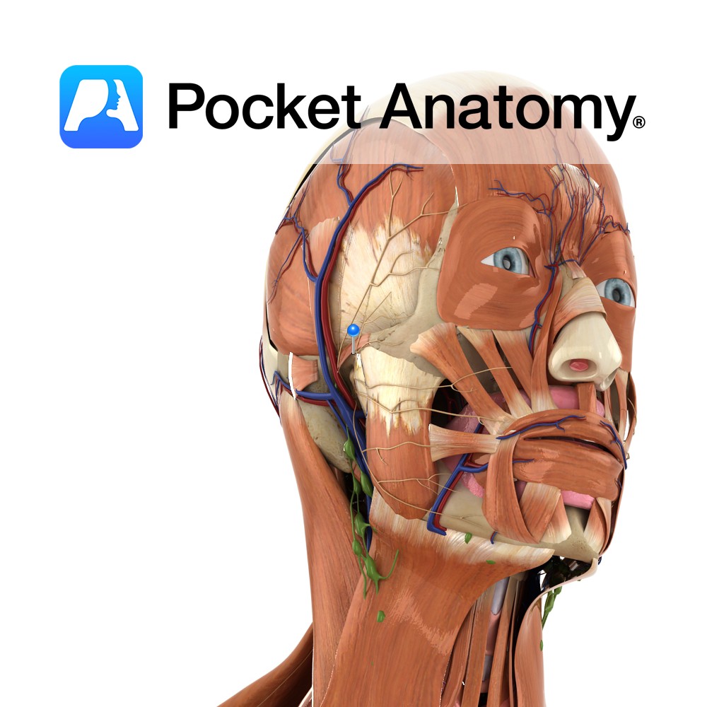
Anatomy Origin: Mastoid process of temporal bone. Insertion: Convexity of the concha of the ear. Key Relations: Lies posterior to the ear on the lateral surface of the head. Functions Retracts and elevates the ear. Supply Nerve Supply: Facial nerve (CN 7) Blood Supply: Posterior auricular artery. Interested in taking our award-winning Pocket Anatomy app
- Published in Pocket Anatomy Pins
Anatomy Origin: Anterior part of the temporal fascia. Insertion: Helix of the ear. Key Relations: Lies on the anterolateral surface of the head. Functions Pulls the ear upward and forward. Supply Nerve Supply: Facial nerve (CN 7) Blood Supply: Superficial temporal artery. Interested in taking our award-winning Pocket Anatomy app for a test drive?
- Published in Pocket Anatomy Pins
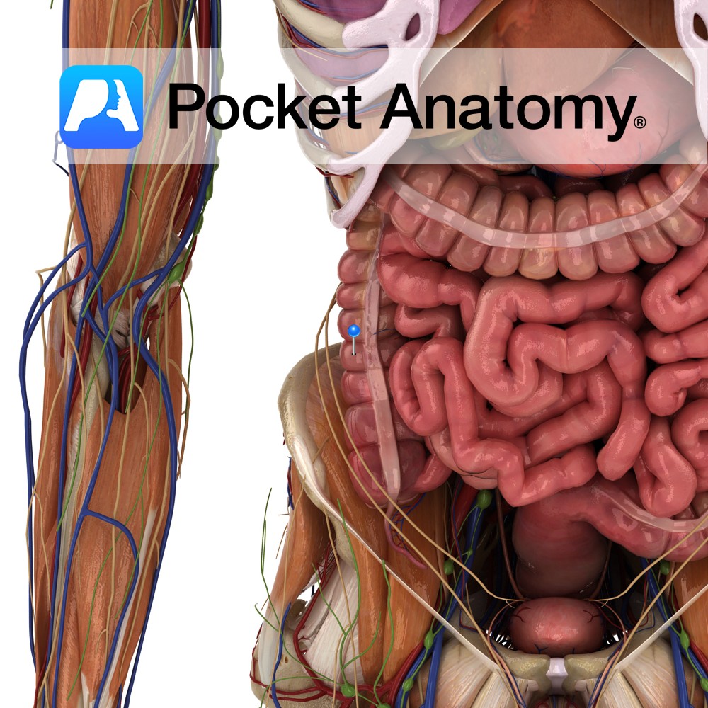
Anatomy Continuous with cecum, extends up right side of abdomen, extra- peritoneally, crossing iliacus and quadratus lumborum and aponeurosis of transversus abdominis, to colic impression under right lobe liver, lateral to gall bladder, where it forms hepatic (right colic) flexure by bending anteriorly and medially (to left) and continuing as the transverse colon. Interested in
- Published in Pocket Anatomy Pins
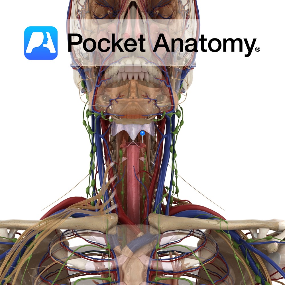
Anatomy Pyramid shaped cartilages sitting on top of the cricoid cartilage. The base is concave and articulates with the slopping articular facet on the superior lateral surface of the lamina of the cricoid cartilage. The anterior angle of the base is extended out into a vocal process. The apex articulates with a corniculate cartilage. Both
- Published in Pocket Anatomy Pins
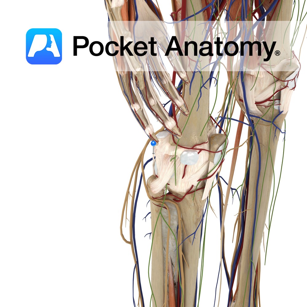
Anatomy An extrinsic ligament of the knee joint. The ligament is Y-shaped and extends from the fibular head. One slip going to the intercondylar area of the tibia. The second slip to the lateral epicondyle of the femur, where it blends with the lateral head of the gastrocnemius. Functions One of the ligaments that help
- Published in Pocket Anatomy Pins
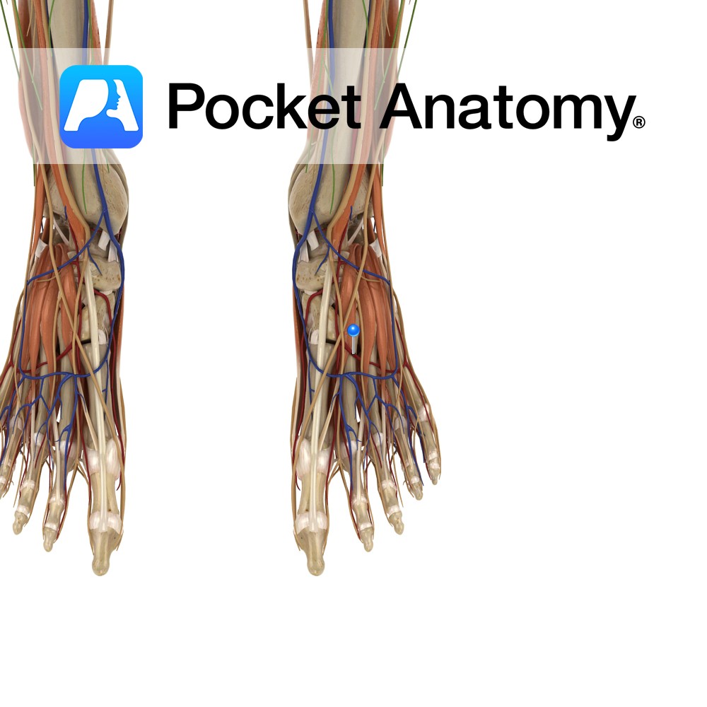
Anatomy Course Branches off the dorsalis pedis artery, where it then proceeds laterally over the dorsal aspect of the metatarsal bones to give off three dorsal metatarsal arteries. Supply Supplies digits two to five. Clinical Not found in all individuals. Interested in taking our award-winning Pocket Anatomy app for a test drive?
- Published in Pocket Anatomy Pins
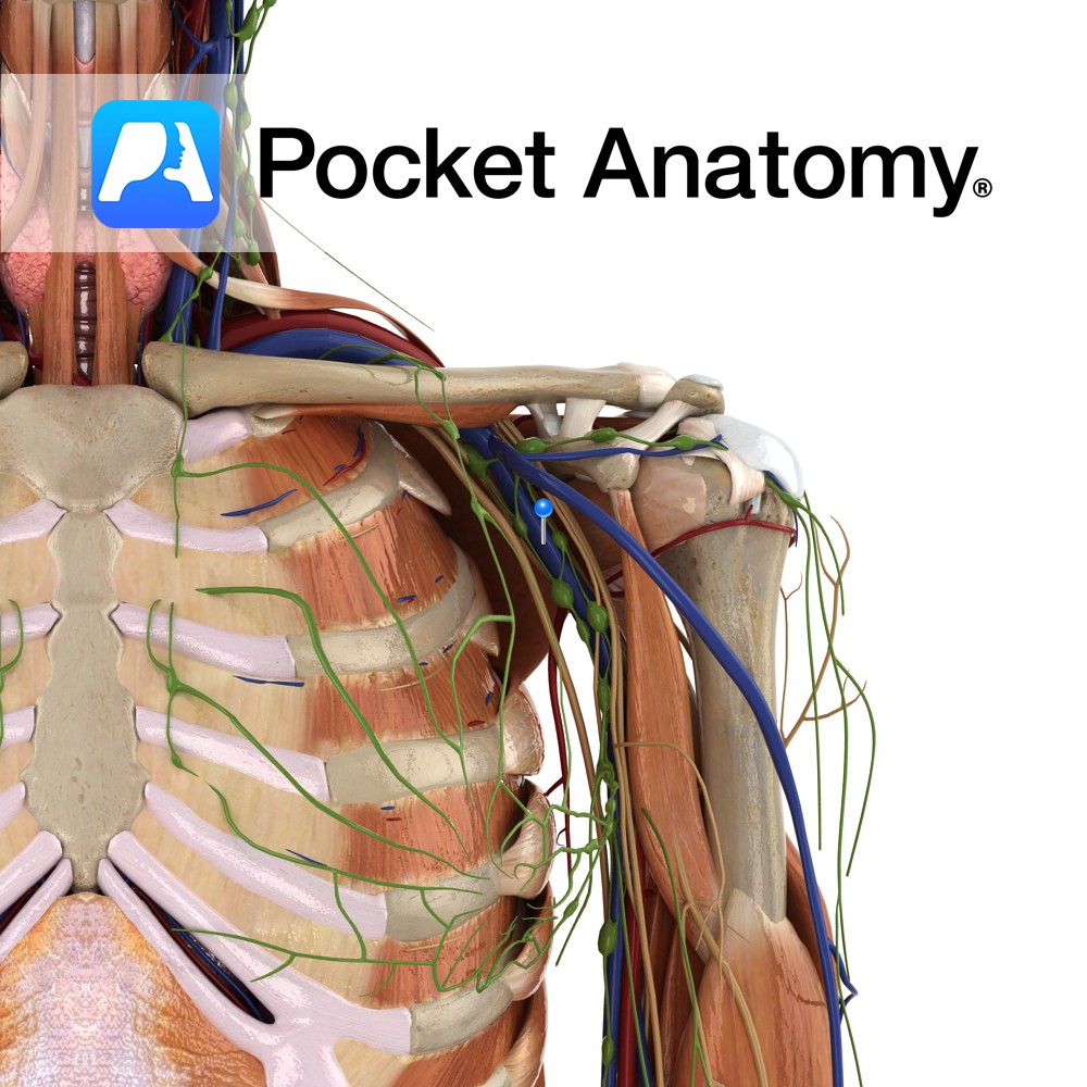
Anatomy Course Continuation of the brachial vein as it passes the teres major muscle. Along with its corresponding artery it travels through the axilla until it reaches the lateral aspect of the first rib, where it becomes the subclavian vein. Drain Receives superficial veins of the area such as the basilic and cephalic veins. Interested
- Published in Pocket Anatomy Pins

