PocketAnatomy® is a registered brand name owned by © eMedia Interactive Ltd, 2009-2022.
iPhone, iPad, iPad Pro and Mac are trademarks of Apple Inc., registered in the U.S. and other countries. App Store is a service mark of Apple Inc.
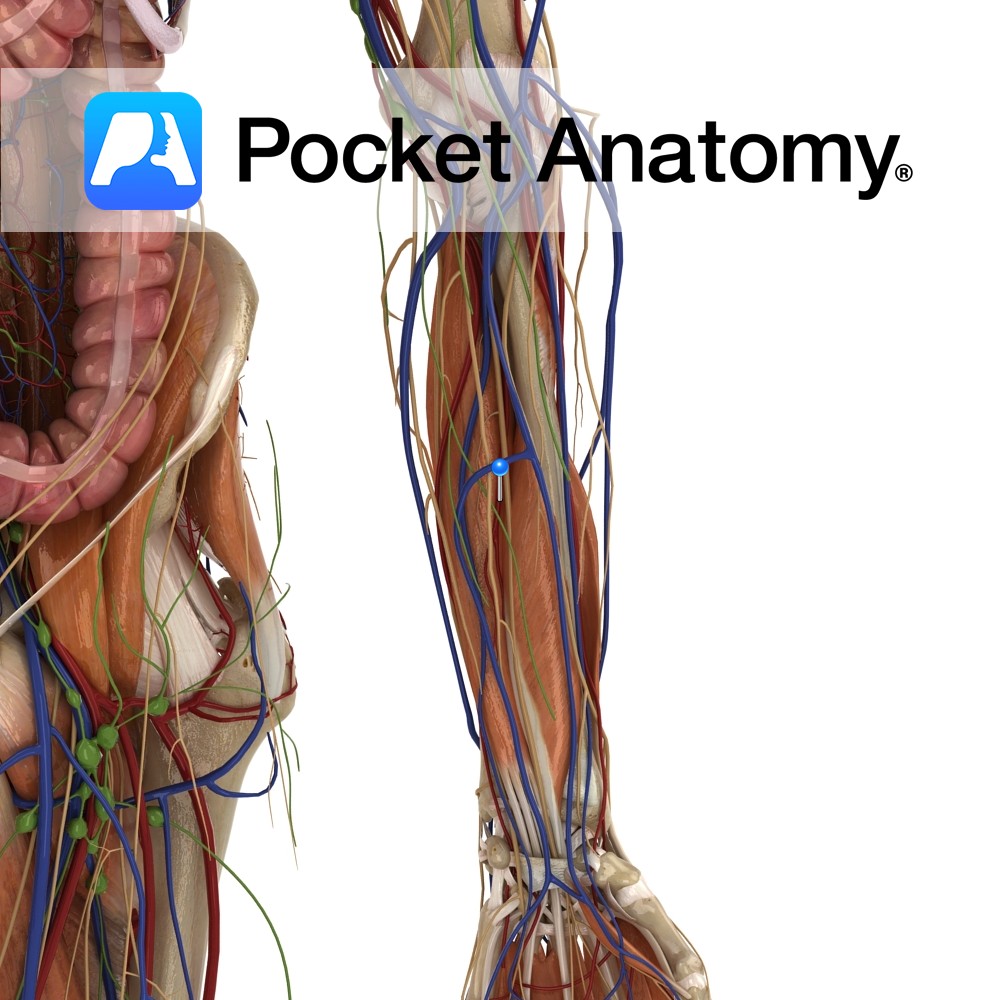
Anatomy Course Branch of the common interosseous artery that originates from the ulnar artery. Rests upon the interosseous membrane of the forearm in the anterior compartment. Branches of the artery pierce the interosseous membrane where it then descends with the dorsal interosseous nerve to the wrist. Deep branches anastamose with the branches of the posterior
- Published in Pocket Anatomy Pins
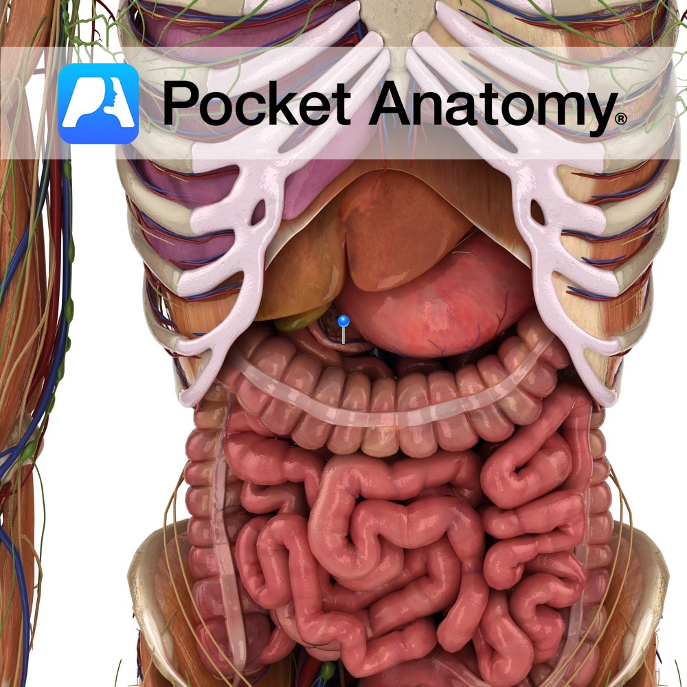
Anatomy Course Travels with its associated artery on the border of the lower duodenum. Drain Drains the pancreas and portions of the duodenum. Interested in taking our award-winning Pocket Anatomy app for a test drive?
- Published in Pocket Anatomy Pins
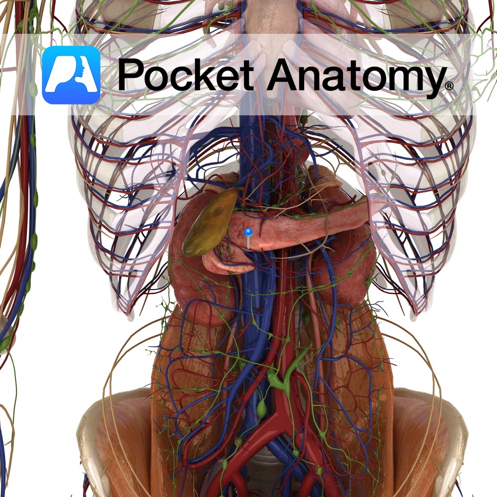
Anatomy Course Branch of the pancreaticoduodenal artery which itself originates from the gastroduodenal artery. Anastamoses with branches of the splenic artery. Supply Responsible for supplying the head of the pancreas as well as the anterior aspect of the duodenum. Interested in taking our award-winning Pocket Anatomy app for a test drive?
- Published in Pocket Anatomy Pins
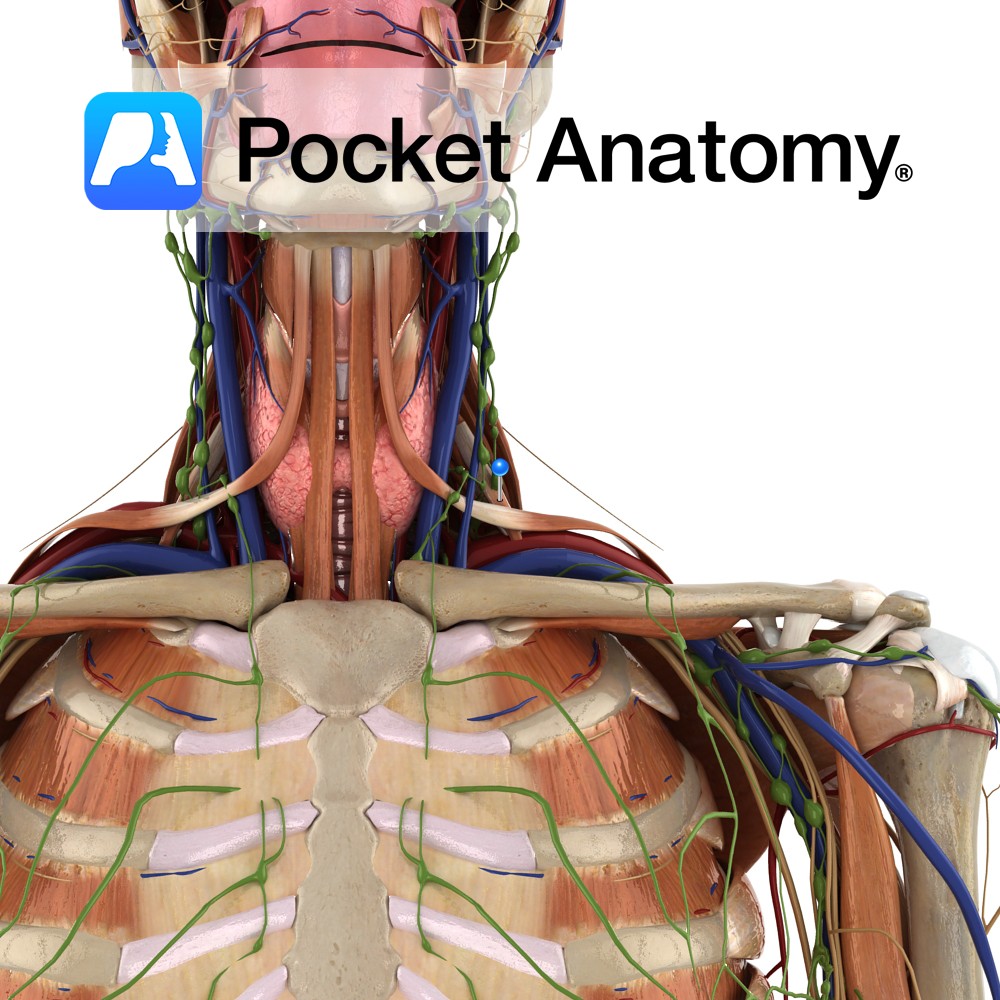
Anatomy Origin: Anterior tubercles of the transverse processes of C3 to C6. Insertion: Scalene tubercle and upper surface of the 1st rib. Key Relations: -Deep to sternocleidomastoid. -The brachial plexus and the subclavian artery pass between the anterior scalene and the middle scalene. -The subclavian vein and phrenic nerve passes anterior to the anterior scalene
- Published in Pocket Anatomy Pins
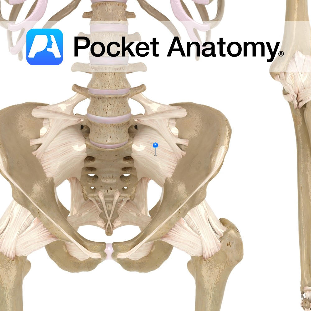
Anatomy A thickening of the fibrous membrane of the sacroiliac joint capsule. It extends from the anterior lateral surface of the sacrum to the articulating margin of the ilium. The ligament covers the anterior and inferior surface of the sacroiliac joint. Functions Stabilizes the sacroiliac joint. Interested in taking our award-winning Pocket Anatomy app for
- Published in Pocket Anatomy Pins
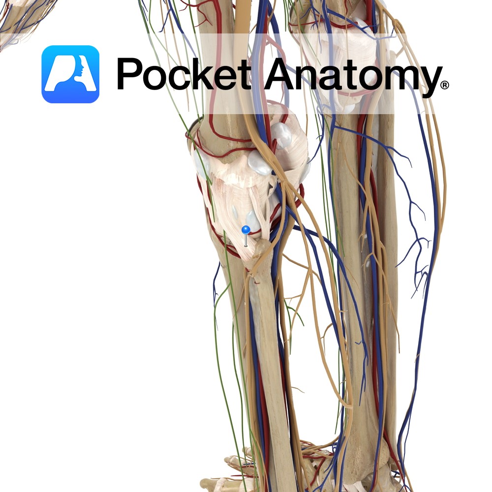
Anatomy Course Branches off the anterior tibial artery immediately after it has crossed the interosseous space. Creates an anastomotic network by connecting arteries on the medial, frontal and lateral aspects of the knee joint. Supply Primarily supplies the knee joint. Interested in taking our award-winning Pocket Anatomy app for a test drive?
- Published in Pocket Anatomy Pins
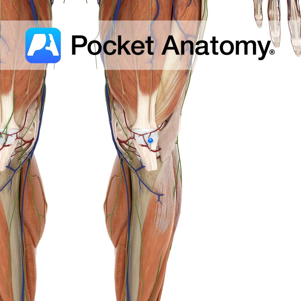
Anatomy The ligament is part of the proximal tibiofibular joint. It attaches to the anterior surface of the head of the fibula to the anterior surface of the head of the tibia, along the oblique line. Functions To reinforce the proximal tibiofibular joint. Interested in taking our award-winning Pocket Anatomy app for a test drive?
- Published in Pocket Anatomy Pins
.jpg)
Anatomy Vermiform (worm-like) blind vestigial sac, 1-8″, arising from 2 cm below ileocecal valve in cecum, right lower quadrant, roughly surface equivalent McBurney’s point (2/3 way from umbilicus to anterior superior iliac spine); base constant but body can lie behind cecum, or in pelvis, or points in between, and can be retroperitoneal. Common site of
- Published in Pocket Anatomy Pins
Anatomy Lowest part of intestine, from lower end sigmoid, down and back about 2.5-4 cms to anus, no peritoneal covering, double sphincter (internal along length and external at anus), supported sling-like by levator ani (thin muscle group of pelvic floor which support its organs and act sphincter-like in maintaining continence and opposing increases in pelvic
- Published in Pocket Anatomy Pins
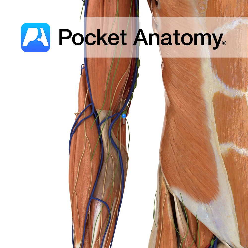
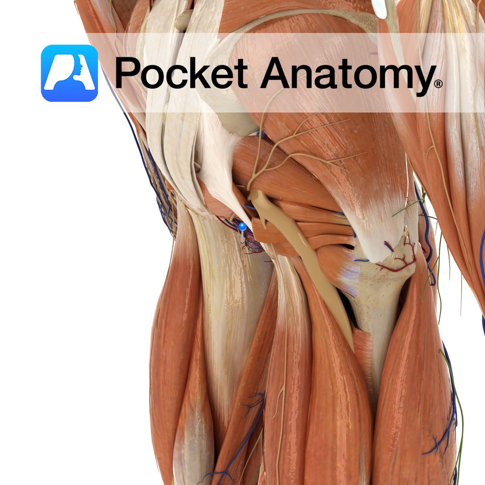
Anatomy Course One of the branches of the ulnar recurrent artery, which assists in the formation of an anastomotic network around the elbow joint. It arises below the elbow joint and rises between the brachialis and pronator teres muscles until it anastomoses with the ulnar collateral arteries. Supply Supplies brachialis and pronator teres muscles as
- Published in Pocket Anatomy Pins