PocketAnatomy® is a registered brand name owned by © eMedia Interactive Ltd, 2009-2022.
iPhone, iPad, iPad Pro and Mac are trademarks of Apple Inc., registered in the U.S. and other countries. App Store is a service mark of Apple Inc.
Anatomy Course A network of nerve fibers running from the spine to neck, axilla and forearm. It is divided into roots (C5, C6, C7, C8, T1), trunks (superior, middle and inferior), divisions (3 anterior and 3 posterior), cords (lateral, posterior and medial), and terminal branches (musculocutaneous, axillary, radial, median and ulnar nerves). Supply Responsible for
- Published in Pocket Anatomy Pins
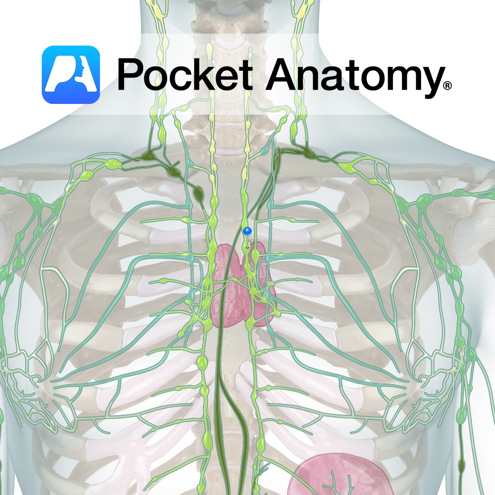
Anatomy Also called bronchial/hilar glands/nodes, situated in pulmonary/lung hilum/root, drain lungs (receive lymph from pulmonary nodes), screen/filter and pass on to tracheobronchial lymph nodes (subgroup of Mediastinal Nodes) and via bronchomediastinal trunk, to Thoracic (Left Lymphatic) Duct. Interested in taking our award-winning Pocket Anatomy app for a test drive?
- Published in Pocket Anatomy Pins
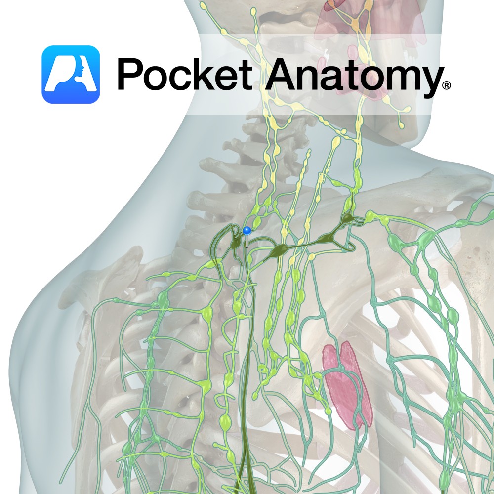
Anatomy One of 4 paired Lymphatic Trunks (Jugular, Subclavian, Bronchomediastinal, Lumbar). Receives lymph from parts of thorax, passes on to Thoracic Duct (L), Right Lymphatic Duct (R) or empties directly into juncture Subclavian and Internal jugular Veins. Clinical Lymph-atic is about transport, Lymph-oid is about T-Lymphocytes. Interested in taking our award-winning Pocket Anatomy app for
- Published in Pocket Anatomy Pins
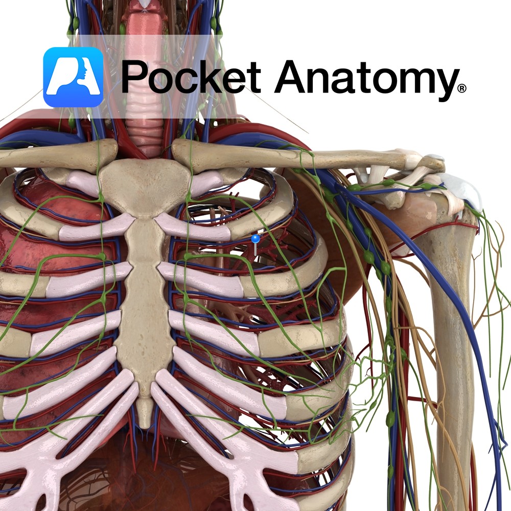
Anatomy That part of airways supported by cartilage, not involved in gas exchange; trachea branches at carina (level sternal angle, T5) – primary bronchi (r, l) – secondary (lobar; 3r, 2l) – tertiary (segmental; 10r, 8- 10l); incrementally down from trachea, as bronchi branch and get smaller, there is more smooth muscle, less hyaline cartilage
- Published in Pocket Anatomy Pins
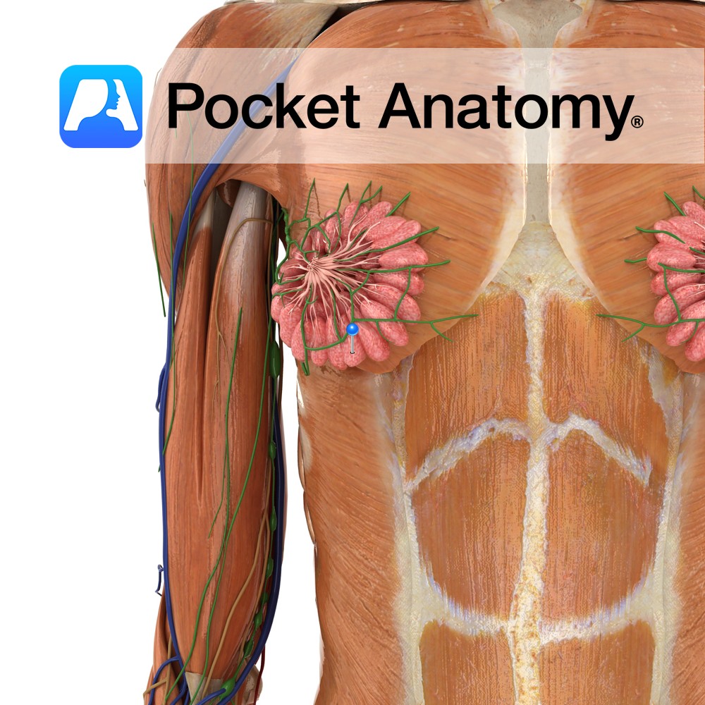
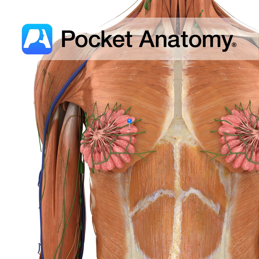
Anatomy Milk-producing component of breast (15-20 in each breast, 10-100 acini/clusters of milk-producing cells each lobe/lobule), drained to the nipple by a duct (breast is modified sweat/apocrine gland, ie an exocrine gland, its secretion being milk), irregularly arranged around the nipple, subcutaneous, composed of fatty tissue in a stroma and supported by ligaments (Cooper’s). Physiology
- Published in Pocket Anatomy Pins
Anatomy Paired skin-covered mammary (milk-producing) gland (modified apocrine/sweat gland) ventral (in front of) pectoralis major muscle on thoracic/chest wall, levels ribs #2 to 6; can extend to axilla at side (sometimes as far as latissimus dorsi at back), up to clavicle, in/medial to sternum, made of adipose/glandular/connective/myoepithelial tissue, comprising 15-20 irregular lobes (each with 10-100
- Published in Pocket Anatomy Pins
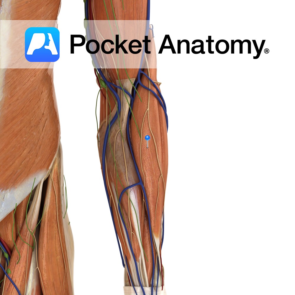
Anatomy Origin: Proximal part of the lateral supracondylar ridge of the humerus and adjacent intermuscular septum. Insertion: Lateral surface of the distal end of the radius. Key Relations: -One of the four muscles of the superficial posterior compartment of the forearm. -Lies anterior to the elbow joint. -Forms the lateral boundary of the cubital fossa.
- Published in Pocket Anatomy Pins
Anatomy Pair of pea-sized exocrine glands below prostate, behind and to side of membranous part of urethra, in front of anus. Duct, 1″, opens into urethra at base penis. Smaller with age. Physiology Secrete salty, thick/viscous alkaline fluid into urethra – during arousal, before ejaculation – which neutralizes acid of residual urine in urethra, and
- Published in Pocket Anatomy Pins
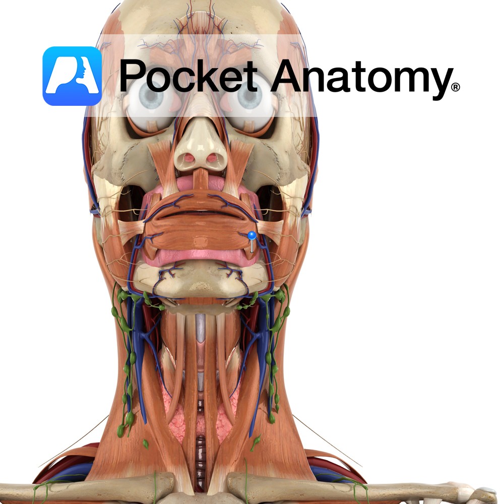
Anatomy Origin: Pterygomandibular raphe, mandible, and alveolar processes of maxilla and mandible. Insertion: Angle of the mouth, some fibres forming deep layers of orbicularis oris. Key Relations: -The parotid duct pierces the muscle opposite the maxillary 3rd molar, before opening into the cavity of the mouth opposite the maxillary 2nd molar. -Often grouped with the
- Published in Pocket Anatomy Pins
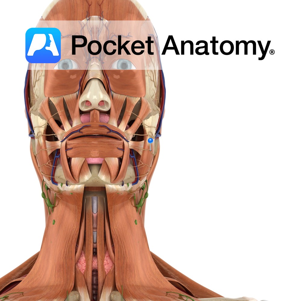
Anatomy Branch of the facial nerve (also known as the seventh cranial nerve). Facial nerve: Has a motor and sensory origin that join together to form the nerve. It passes through the internal auditory meatus through the facial canal and finally exits from the stylomastoid foramen, and into the parotid gland where it divides into
- Published in Pocket Anatomy Pins

.jpg)
