PocketAnatomy® is a registered brand name owned by © eMedia Interactive Ltd, 2009-2022.
iPhone, iPad, iPad Pro and Mac are trademarks of Apple Inc., registered in the U.S. and other countries. App Store is a service mark of Apple Inc.
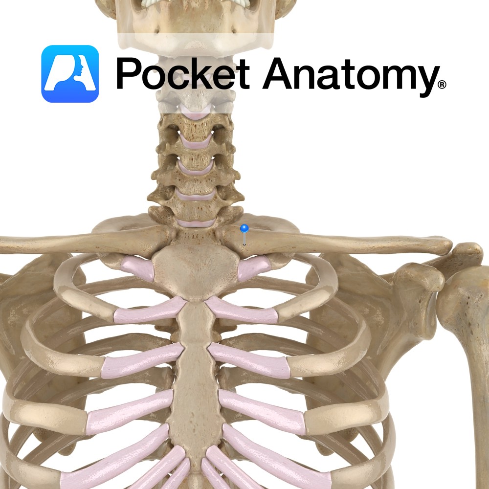
Anatomy Medial end of clavicle (which is a long flat bone, elongated s-shape, convex at medial end, concave at lateral) articulates with manubrium of sternum. Clinical The only horizontal long bone (though ordinarily, no marrow), the clavicle acts as a strut, holding scapula in place and free to move on thoracic wall, which in turn
- Published in Pocket Anatomy Pins
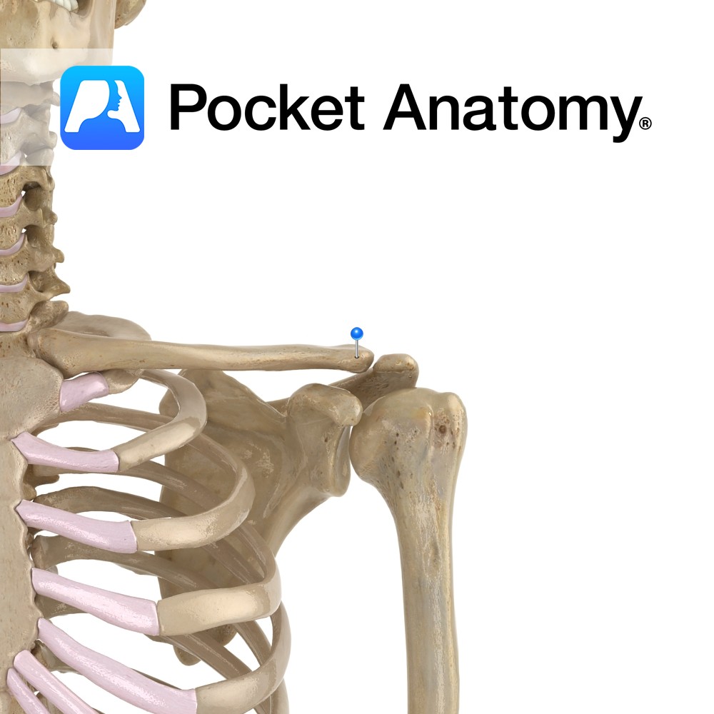
Anatomy Lateral end of clavicle (which is a long flat bone, elongated s-shape, convex at medial end, concave at lateral) articulates with acromial process of scapula. Connected to coracoid process below by coracoclavicular ligament. Clinical Clavicula (Latin); little key. First bone to begin ossification (5-6 weeks in embryo), one of last to finish (21-25 years).
- Published in Pocket Anatomy Pins
.jpg)
Anatomy Course Arises from the third division axillary artery at the inferior border of the subscapularis muscle. In association with the axillary nerve it travels through the quadrangular space. Supply Supplies the glenohumeral joint and the neck of the humerus. Interested in taking our award-winning Pocket Anatomy app for a test drive?
- Published in Pocket Anatomy Pins
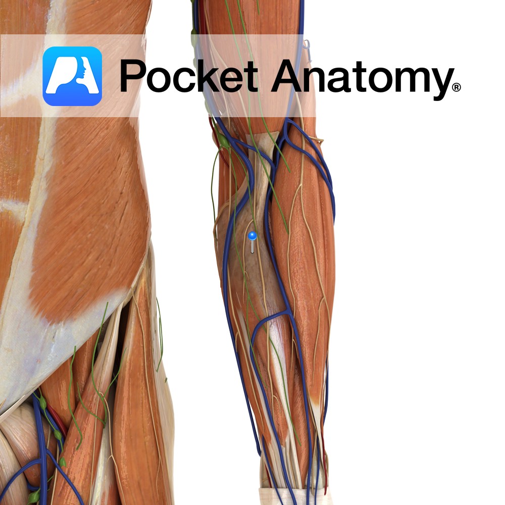
Anatomy As the biceps brachii tendon enters the forearm, the medial portion of the tendon sends off a slip medially. This slip fans out and attaches to the deep fascia of the medial forearm. The fanned out tendon is the aponeurousis. Functions It is one of the distal attachments of the biceps brachii muscle in
- Published in Pocket Anatomy Pins
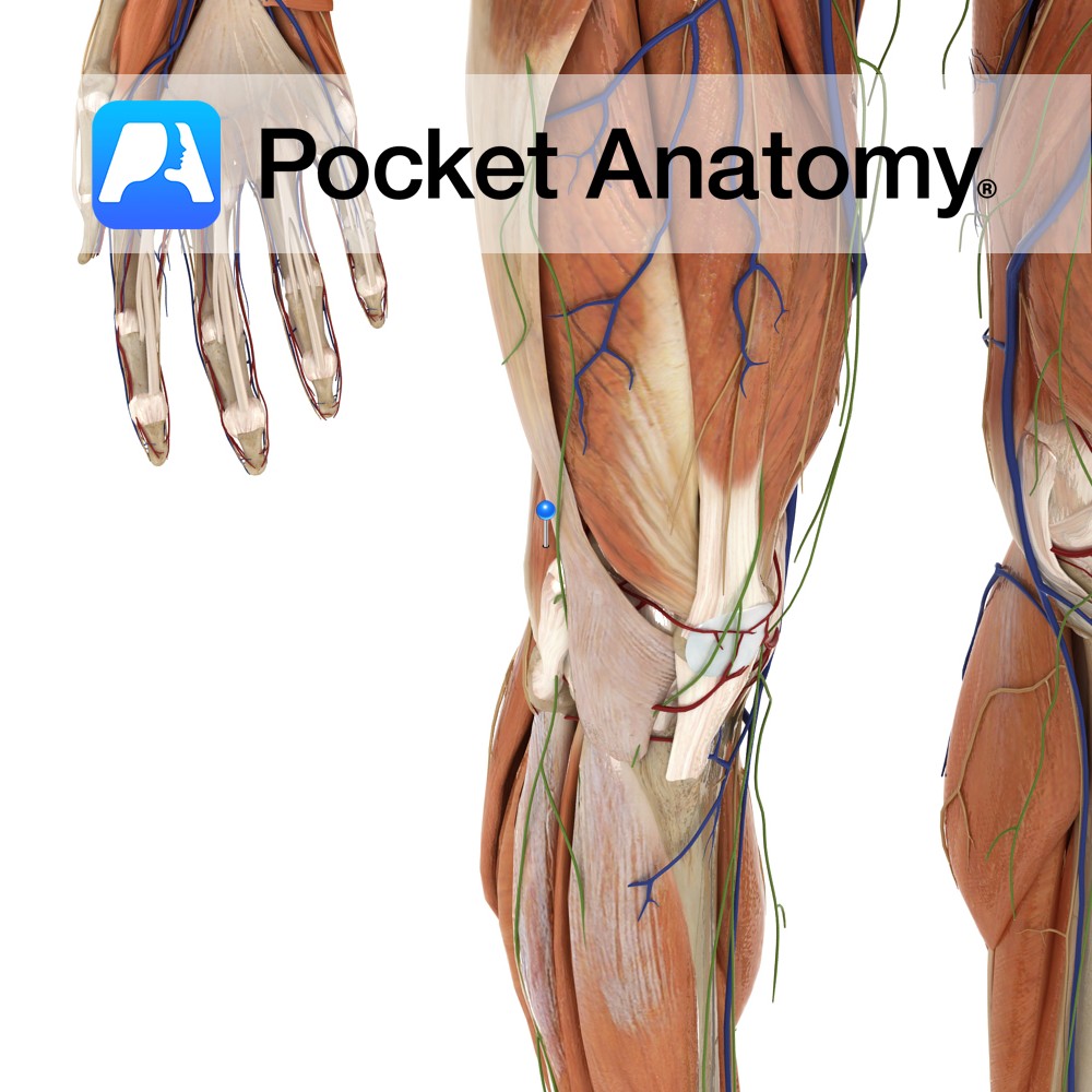
Anatomy Origin: Long head: Superomedial area of ischial tuberosity (with semitendinosus). Short head: Lateral lip of linea aspera and upper lateral supracondylar line of femur and lateral intermuscular septum. Insertion: The two heads insert by a common tendon into the lateral surface of head of fibula. Key Relations: -One of the three muscles of the
- Published in Pocket Anatomy Pins
.jpg)
Anatomy Origin: Short head: Coracoid process of the scapula. Long head: Tendon from supraglenoid tubercle of the scapula. Insertion: By common tendon into radial tuberosity and also forms bicipital aponeurosis on medial aspect of forearm. Key Relations: -The brachial artery and the median nerve run together on the medial border until the distal one third
- Published in Pocket Anatomy Pins
.jpg)
Anatomy Origin: Long head: Tendon from supraglenoid tubercle of the scapula. Short head: Coracoid process of the scapula. Insertion: By common tendon into radial tuberosity and also forms bicipital aponeurosis on medial aspect of forearm. Key Relations: -The brachial artery and the median nerve run together on the medial border until the distal one third
- Published in Pocket Anatomy Pins
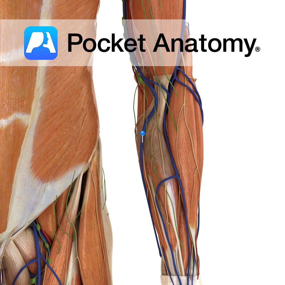
Anatomy Course Commences on the ulnar side of the dorsum of the hand where it then takes a superficial path up the forearm resting on top of the superficial muscles. As it nears the elbow, it usually creates connections with the cephalic vein before finally joining the axillary vein in upper arm. Drain Drains portions
- Published in Pocket Anatomy Pins
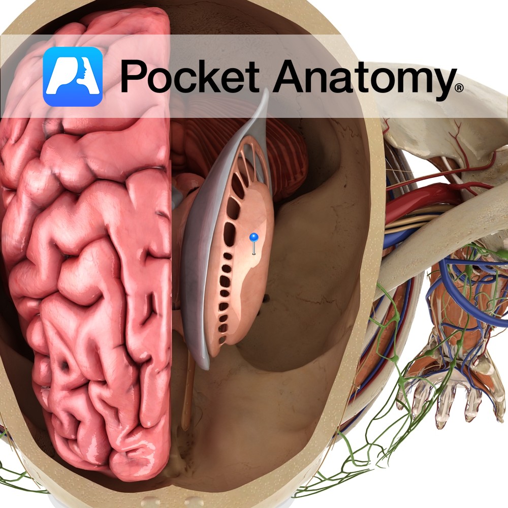
Anatomy The basal ganglia is a collection of nuclear masses located within each cerebral hemisphere. The most prominent bodies are the caudate nucleus, putamen and globus pallidus. These nuclei are collectively referred to as the corpus striatum. Blood Supply: Supplied by branches of the internal carotid, anterior cerebral and middle cerebral arteries. Functions The basal
- Published in Pocket Anatomy Pins
.jpg)
Anatomy Course A network of nerve fibers running from the spine to neck, axilla and forearm. It is divided into roots (C5, C6, C7, C8, T1), trunks (superior, middle and inferior), divisions (3 anterior and 3 posterior), cords (lateral, posterior and medial), and terminal branches (musculocutaneous, axillary, radial, median and ulnar nerves). Supply Responsible for
- Published in Pocket Anatomy Pins