PocketAnatomy® is a registered brand name owned by © eMedia Interactive Ltd, 2009-2022.
iPhone, iPad, iPad Pro and Mac are trademarks of Apple Inc., registered in the U.S. and other countries. App Store is a service mark of Apple Inc.
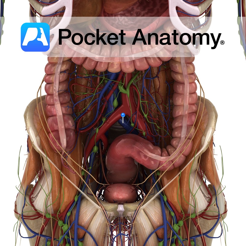
Anatomy Course Formed by the fusion of the external and internal iliac veins at the brim of the pelvis. Travel briefly before the two common iliac veins fuse together and form the inferior vena cava at the level of L5. Drain Receives the deoxygenated blood of the lower limb and pelvis. Clinical The right iliac
- Published in Pocket Anatomy Pins
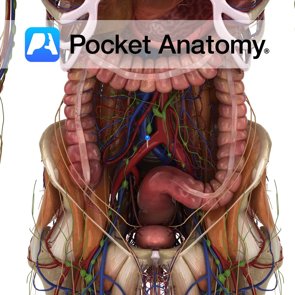
Anatomy Course Large branches of the bifurcation of the abdominal aorta at the level of L4. Travels along the inferior edge of the psoas muscles. Supply Responsible for the supply of the pelvis and the lower limb via the femoral artery. Clinical Common area for stenosis due to atherosclerosis; this is more common in the
- Published in Pocket Anatomy Pins
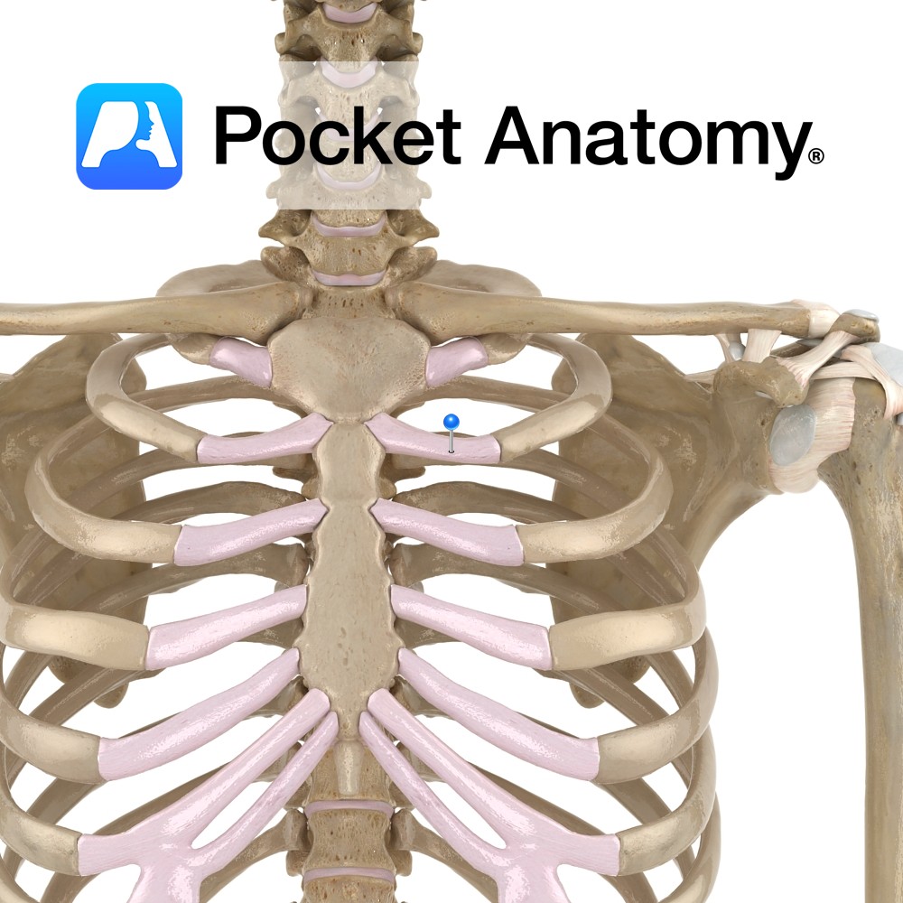
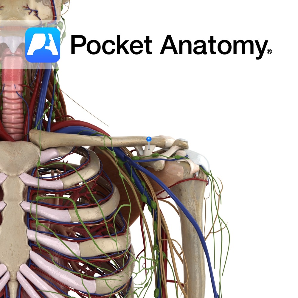
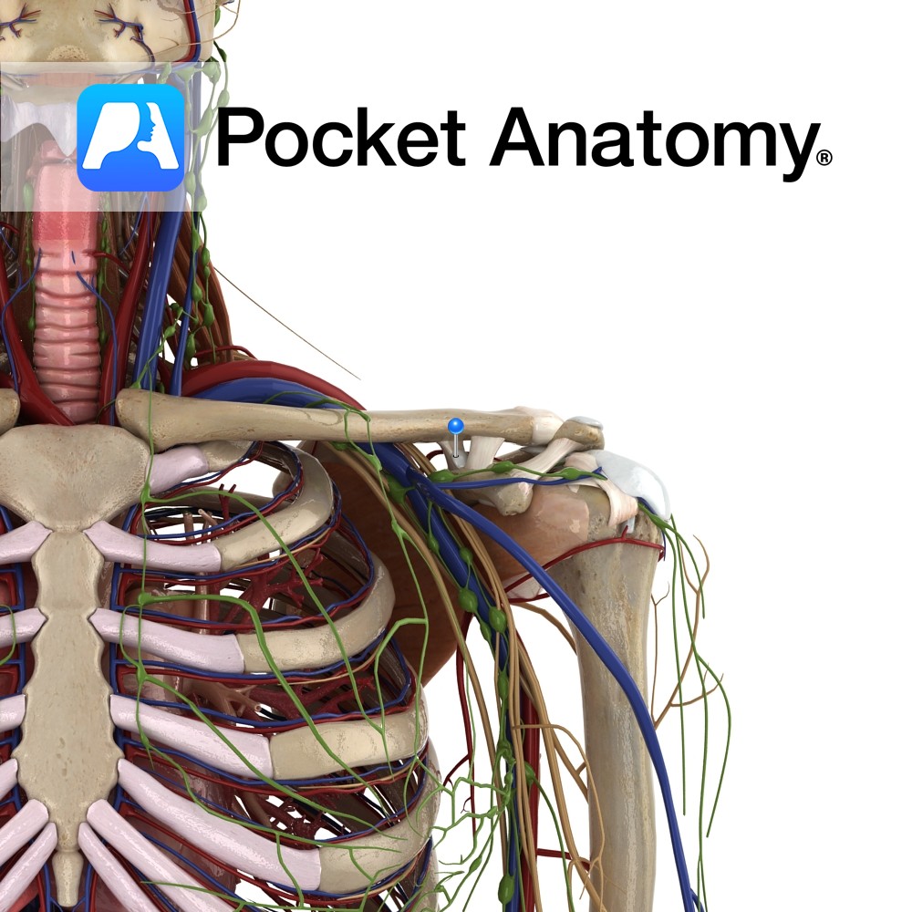
Anatomy Cartilaginous forward/anterior/sternal prolongations of the ribs, articulating with the sternum, giving the thorax elasticity with strength. Clinical Hyaline cartilage is found on articular surfaces of synovial joints (as a thin covering layer with a smooth surface), providing resilience. Also found in nasal septum, larynx, trachea and bronchi, where it provides elasticity. Costal cartilage has
- Published in Pocket Anatomy Pins
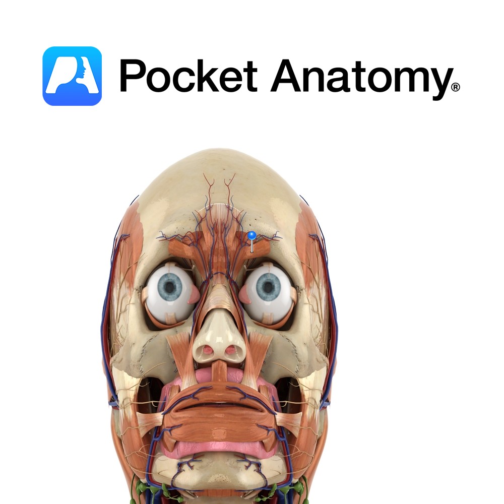
Anatomy Origin: Medial end of superciliary arch. Insertion: Skin of medial half of eyebrow. Key Relations: Lies deep to orbicularis oculi and the eyebrows. Functions Draws eyebrows medially and downwards. e.g. when frowning.. Supply Nerve Supply: Temporal branch of the facial nerve (CN 7). Blood Supply: Opthalmic artery. Interested in taking our award-winning Pocket Anatomy
- Published in Pocket Anatomy Pins
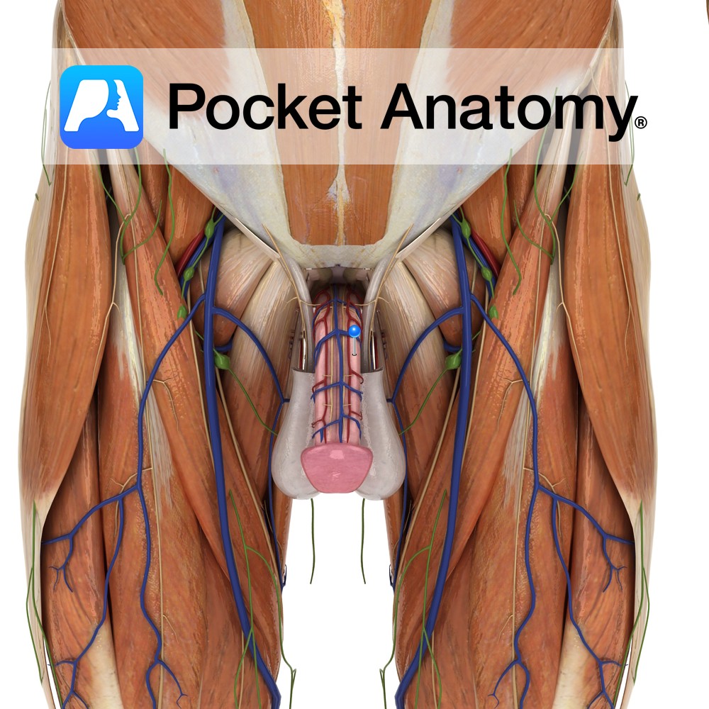
Anatomy There are 3 cylindrical strips of expandable/inflatable/erectile tissue along length of penis; 2 corpus cavernosum side by side and dorsal to 1 (smaller) corpus spongiosum (central and ventral, expanded at proximal end into bulb and distal into glans, and through which urethra courses). Corpus cavernosum starts at pubic bone, left and right end by
- Published in Pocket Anatomy Pins
Anatomy The coracohumeral ligament attaches from the lateral surface of the coracoid process to blend with the supraspinatus tendon, and attach to the superior surface of the greater tuberosity of the humerus. Functions Helps to keep the head of the humerus in contact with glenoid fossa of the scapula. Provides support and stability to the
- Published in Pocket Anatomy Pins
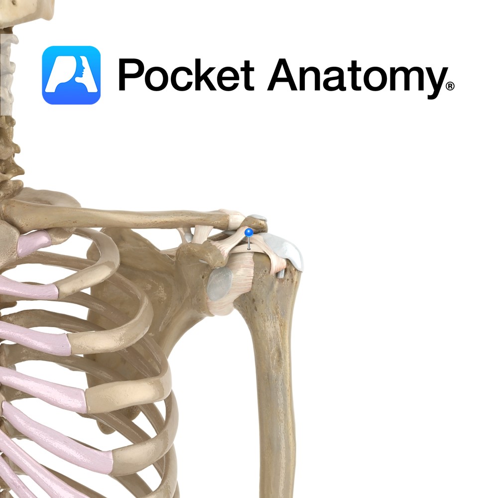
Anatomy The trapezoid (strap-like) ligament attaches along the trapezoid line of the clavical to the posterior aspect of the coracoid process. Functions This ligament is not directly related to the acromioclavicular joint but is a strong accessory ligament. It maintains the position of the clavical on the acromion and also gives weight bearing support to
- Published in Pocket Anatomy Pins
Anatomy The conoid (cord-like) ligament attaches from the conoid tubercle of the clavicle to the posterior aspect of the coracoid process. Functions This ligament is not directly related to the acromioclavicular joint but is a strong accessory ligament. It maintains the position of the clavical on the acromion and also gives weight bearing support to
- Published in Pocket Anatomy Pins
.jpg)
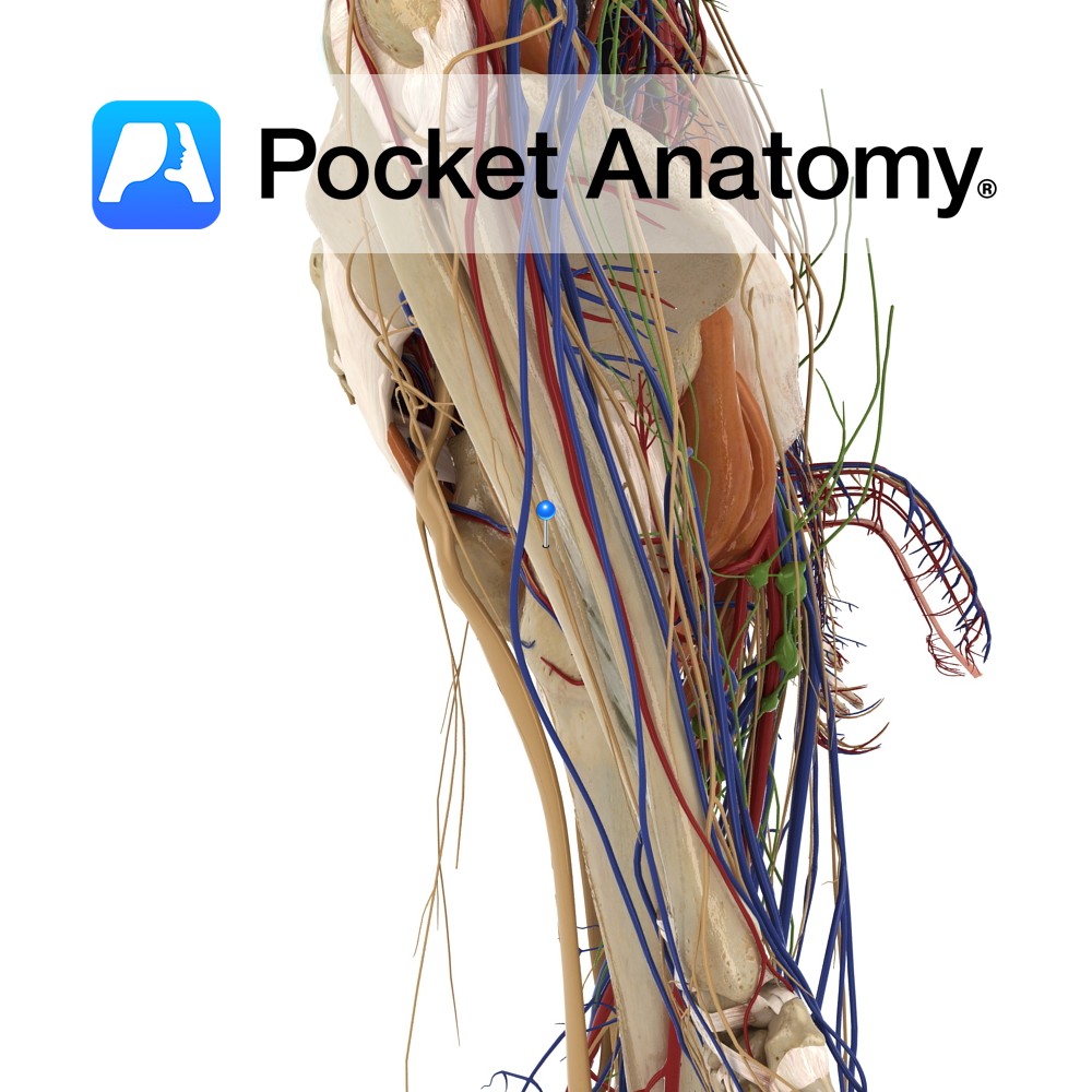
Anatomy Course Branches off the lateral aspect of the femoral artery in the femoral triangle. It then descends between the pectineus and the adductor longus. Supply Supplies the majority of the thigh. Interested in taking our award-winning Pocket Anatomy app for a test drive?
- Published in Pocket Anatomy Pins
Anatomy Course As the name suggests it is a branch of the radial nerve which travels posteriorly to lateral aspect of the radius in between the supinator muscle. It continues a downwards path until it reaches approximately the middle of the forearm, where it continues as the posterior interosseous nerve. Supply Motor innervation to the
- Published in Pocket Anatomy Pins