PocketAnatomy® is a registered brand name owned by © eMedia Interactive Ltd, 2009-2022.
iPhone, iPad, iPad Pro and Mac are trademarks of Apple Inc., registered in the U.S. and other countries. App Store is a service mark of Apple Inc.
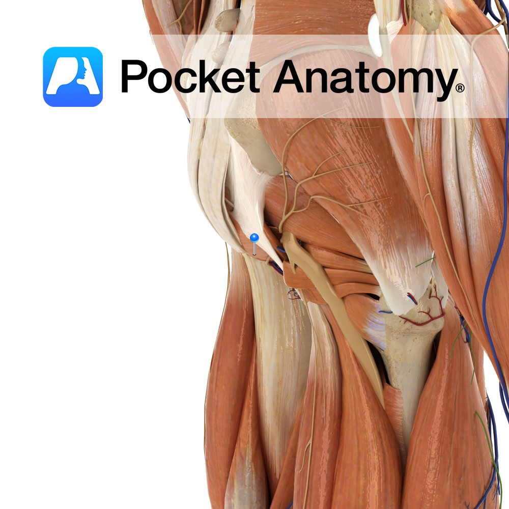
Anatomy Course Branch of the genicular artery that pierces the adductor canal, where it then travels to the medial aspect of the knee with the saphenous nerve. Supply Anastomotic network around the knee. Interested in taking our award-winning Pocket Anatomy app for a test drive?
- Published in Pocket Anatomy Pins
Anatomy Course Branch of the descending genicular artery, which then proceeds to descend on the adductor magnus tendon until it reaches the medial aspect of the knee. Supply Anastomotic network around the knee. Interested in taking our award-winning Pocket Anatomy app for a test drive?
- Published in Pocket Anatomy Pins
Anatomy Articulates up with middle phalanx at 5th DIP. Interested in taking our award-winning Pocket Anatomy app for a test drive?
- Published in Pocket Anatomy Pins
Anatomy Articulates up with middle phalanx at 3rd DIP. Interested in taking our award-winning Pocket Anatomy app for a test drive?
- Published in Pocket Anatomy Pins
Anatomy The more distal of the 2 thumb phalanges, connected above by the intermediate interphalangeal joint. Interested in taking our award-winning Pocket Anatomy app for a test drive?
- Published in Pocket Anatomy Pins
Anatomy Articulates up with 5th middle phalanx. Interested in taking our award-winning Pocket Anatomy app for a test drive?
- Published in Pocket Anatomy Pins
Anatomy Articulates up with 3rd middle phalanx. Interested in taking our award-winning Pocket Anatomy app for a test drive?
- Published in Pocket Anatomy Pins
Anatomy Articulates proximally with 1st proximal phalanx. Interested in taking our award-winning Pocket Anatomy app for a test drive?
- Published in Pocket Anatomy Pins
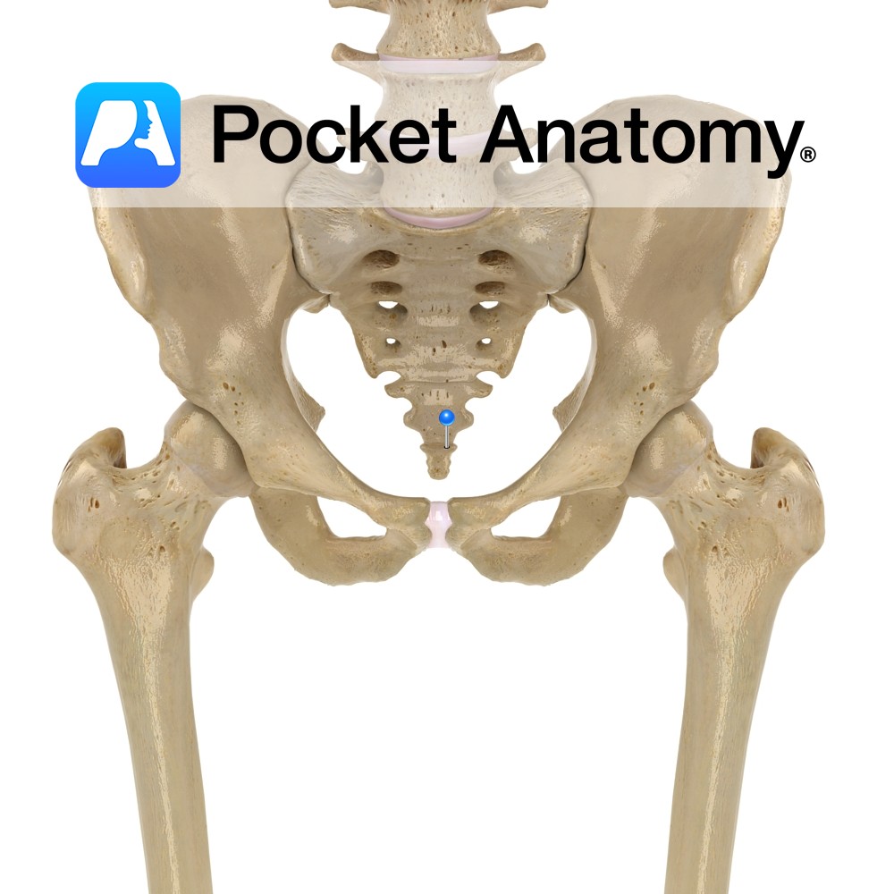
Anatomy Tailbone. Lowest section of spine, made up of 3-5 small vertebrae, often fused. Transition top to bottom of coccyx, from rudimentary to absent pedicles, laminae, transverse and spinous processes. Clinical Articulates with Sacrum above; limited movement. Vignette From Greek for cuckoo, as viewed from side, resembles its beak. Vestigial tail. Interested in taking our
- Published in Pocket Anatomy Pins
Anatomy Origin: Ischial spine and the pelvic surface of the sacrospinous ligament. Insertion: Lateral margin of the coccyx and related border of the sacrum. Key Relations: -Coccygeus overlies the sacrospinous ligament. -Contributes to the formation of the posterior part of the pelvic diaphragm in association with the levator ani muscles- iliococcygeus (for more information see
- Published in Pocket Anatomy Pins

.jpg)
.jpg)
.jpg)
.jpg)
.jpg)
.jpg)
.jpg)
.jpg)

