PocketAnatomy® is a registered brand name owned by © eMedia Interactive Ltd, 2009-2022.
iPhone, iPad, iPad Pro and Mac are trademarks of Apple Inc., registered in the U.S. and other countries. App Store is a service mark of Apple Inc.
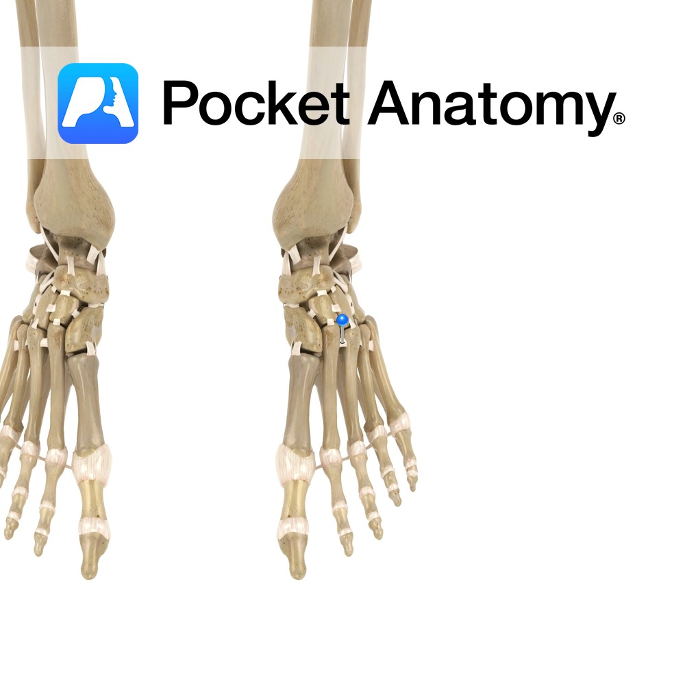
Anatomy A series of ligaments that attach from the dorsal surface of one metatarsal to the dorsal surface of the adjacent metatarsal. Situated at the proximal ends of the metatarsal bones. Functions Stabilizers of the metatarsal joints. Interested in taking our award-winning Pocket Anatomy app for a test drive?
- Published in Pocket Anatomy Pins
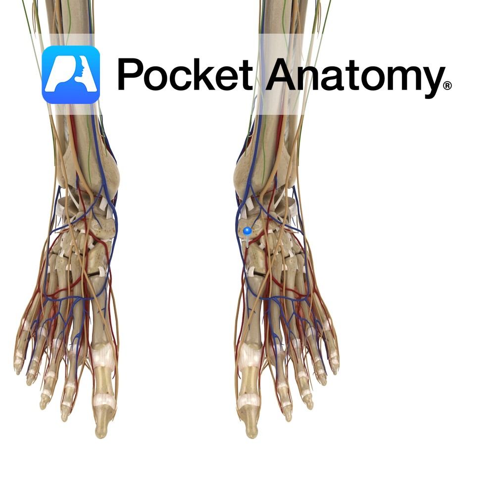
Anatomy Course First dorsal metatarsal artery is a direct branch of the dorsalis pedis artery. The second, third and fourth metatarsal arteries originate from the arcuate artery. Supply Together they supply the digits of the foot. Interested in taking our award-winning Pocket Anatomy app for a test drive?
- Published in Pocket Anatomy Pins
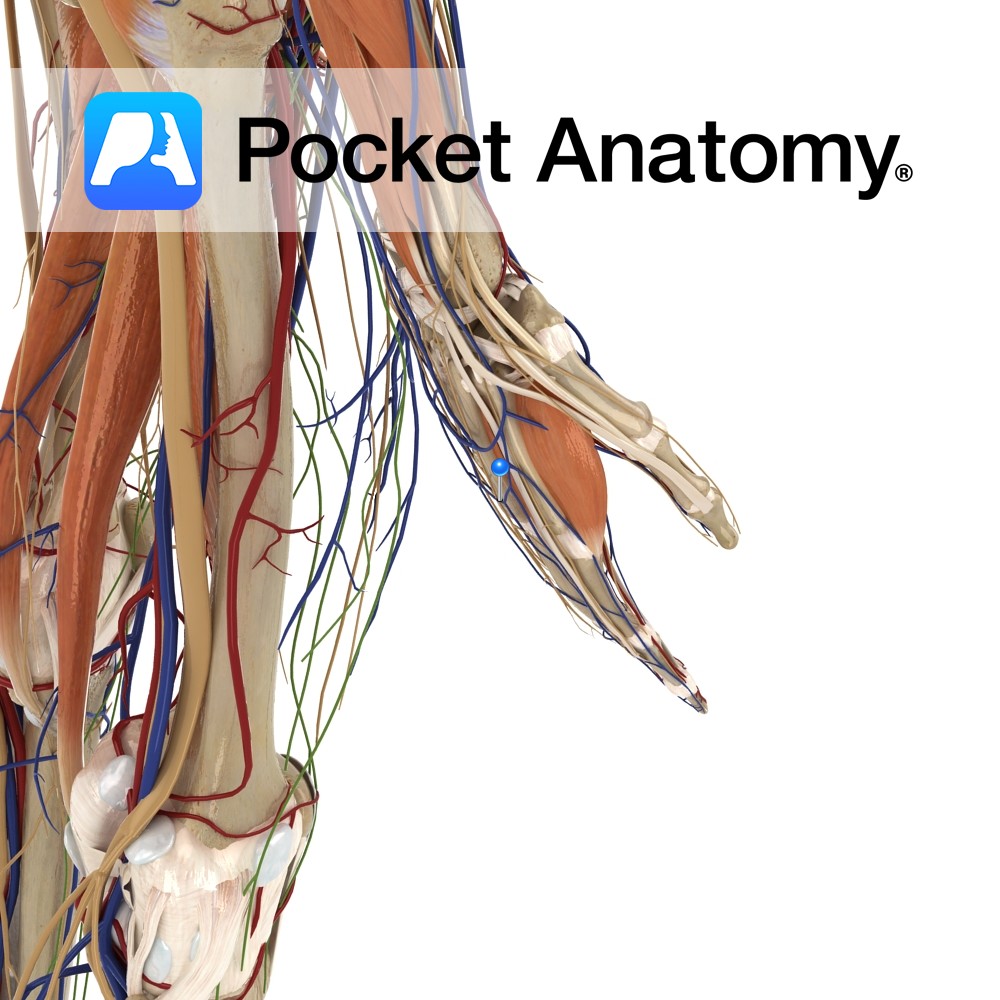
Anatomy Course Arise from the dorsal digital veins, which form three dorsal metacarpal veins. These travel briefly before creating a complex network on the dorsum of the hand. Drain Drain the dorsum of the hand. Interested in taking our award-winning Pocket Anatomy app for a test drive?
- Published in Pocket Anatomy Pins
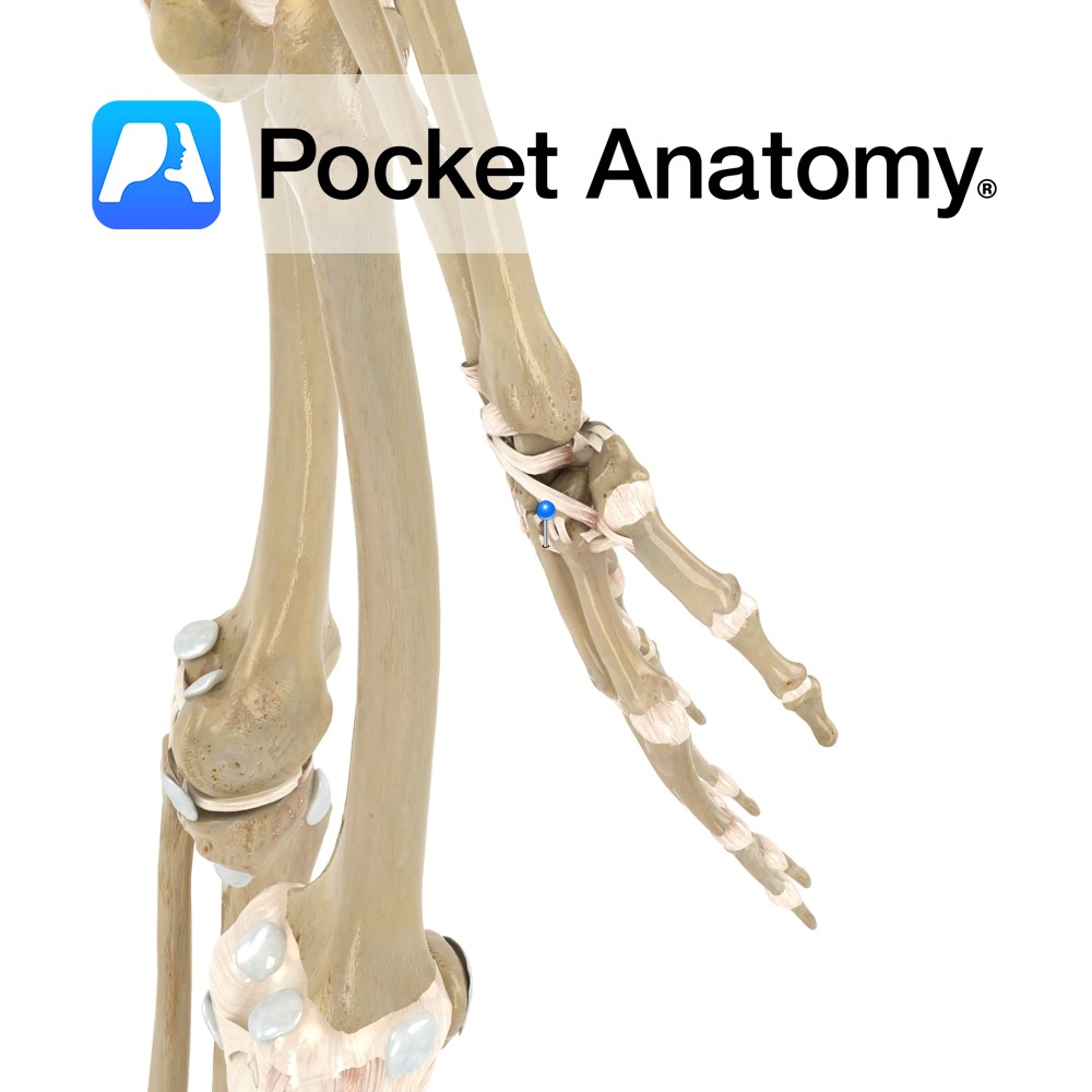
Anatomy Thick bands of connective tissue attaching from the bases of the two to five metacarpal bones. Functions By linking metacarpal bones two to five, they help to form the skeletal framework of the palm. Interested in taking our award-winning Pocket Anatomy app for a test drive?
- Published in Pocket Anatomy Pins
.jpg)
Anatomy Origin: Bipennate muscles that arise by two heads from adjacent sides of posterior aspect of the metacarpals. Insertion: Extensor hood and base of proximal phalanges of the index, middle and ring fingers. (It is the middle digit that has two dorsal interossei inserting onto it.) Key relations: The radial artery (after passing between the
- Published in Pocket Anatomy Pins
.jpg)
Anatomy Origin: Sides of the adjacent metatarsals. Insertion: Bases of proximal phalanges of 2nd, 3rd and 4th toes and dorsal expansions of the extensor digitorum longus tendons. Key relations: The dorsal interossei lie dorsal to the plantar interossei and together they form the fourth layer of the plantar muscles of the foot. Also, see view
- Published in Pocket Anatomy Pins
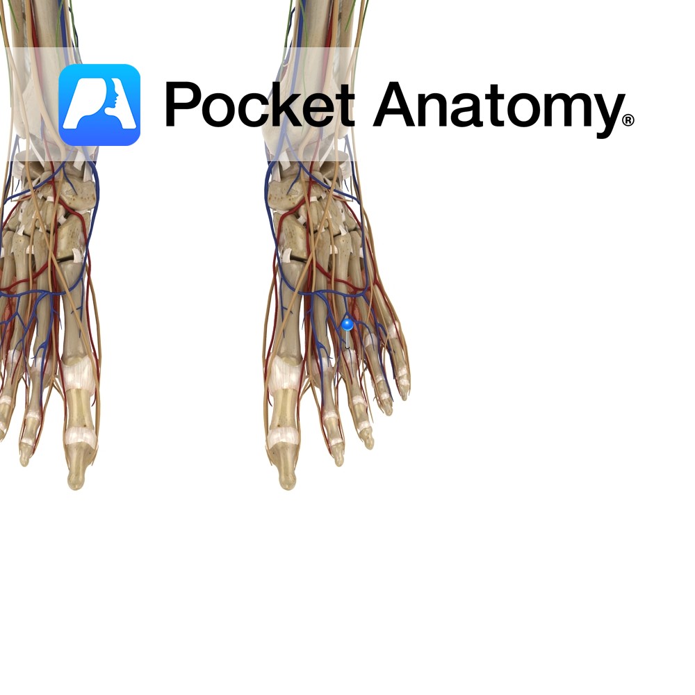
Anatomy Course On the dorsum of the foot they receive the intercapitular veins. They travel briefly to join the plantar cutaneous venous arch. Drain Drains the dorsal surface of the digits of the foot. Interested in taking our award-winning Pocket Anatomy app for a test drive?
- Published in Pocket Anatomy Pins
.jpg)
Motion The elbow joint is a uniaxial compound synovial hinge joint. The elbow joint actually includes 2 joints the humeroulnar joint and the humeroradial joint. The capitulum of the humerus articulates with the head of the radius, and the trochlea of the humerus articulates with the trochlear notch of the ulna. Together these articulations make
- Published in Pocket Anatomy Pins
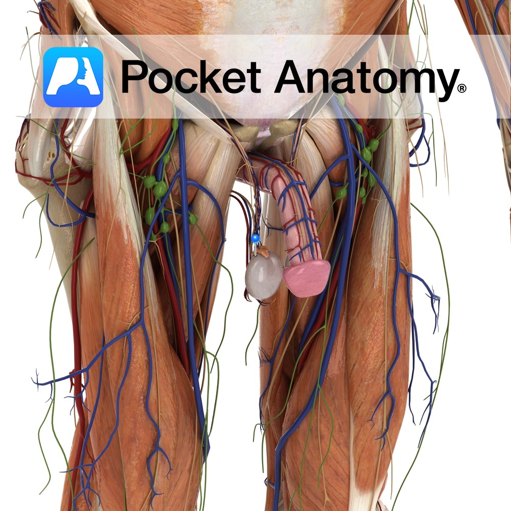
Anatomy 15-20 muscular ducts which connect rete testis to epididymis, accounting for 1/3 volume head of epididymis, concentrating spermatozoa by water reabsorption and moving them on. Interested in taking our award-winning Pocket Anatomy app for a test drive?
- Published in Pocket Anatomy Pins

Anatomy The dura mater is the strong, thick, outermost layer of the meninges attached to the periosteum of the skull except where it runs long the skull base or is reflected along the skull vault. Venous sinuses run inside spaces within the dura mater created by separation of the dura mater from the periosteum at
- Published in Pocket Anatomy Pins