PocketAnatomy® is a registered brand name owned by © eMedia Interactive Ltd, 2009-2022.
iPhone, iPad, iPad Pro and Mac are trademarks of Apple Inc., registered in the U.S. and other countries. App Store is a service mark of Apple Inc.
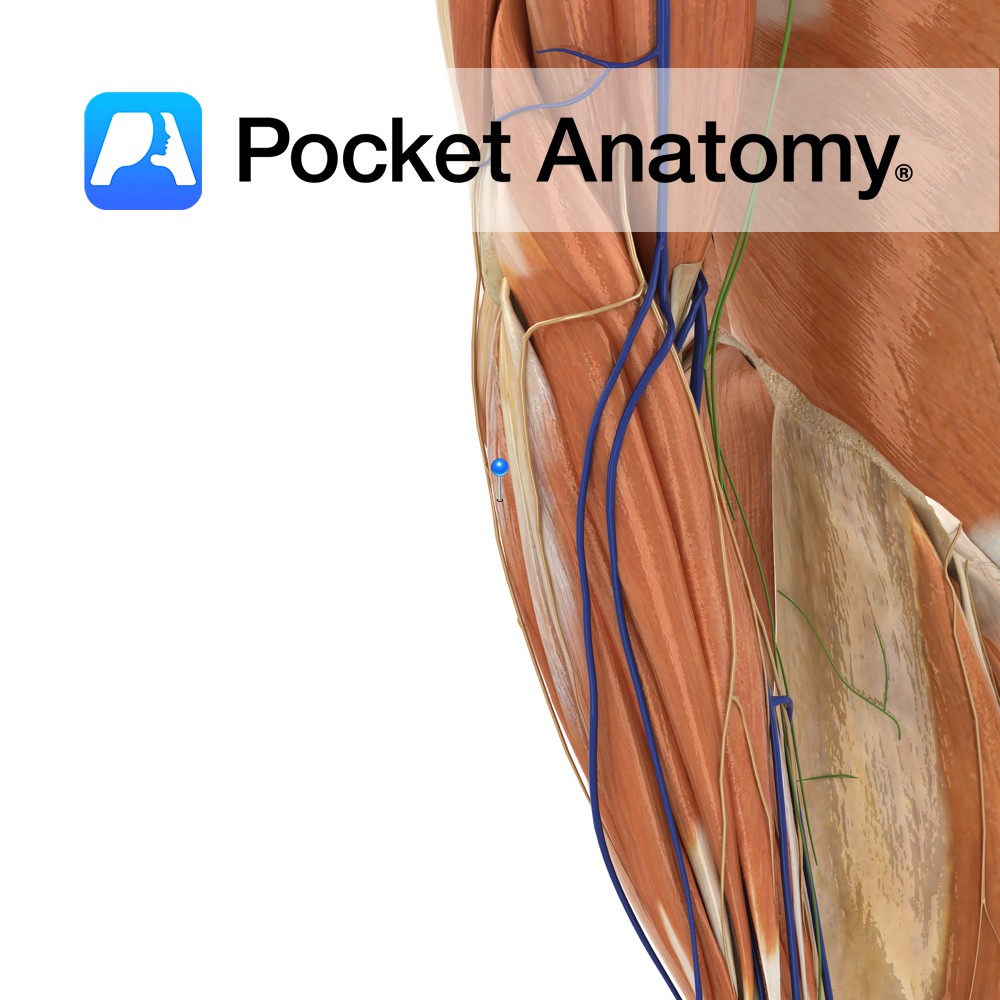
Anatomy Origin: First head: Lateral epicondyle of humerus via the common extensor tendon. Second head: Posterior border of ulna. Insertion: Medial aspect of base of fifth metacarpal Key Relations: One of the four muscles in the superficial posterior compartment of the forearm. Functions -Extends the hand at the wrist joint. -Adducts the hand at the
- Published in Pocket Anatomy Pins
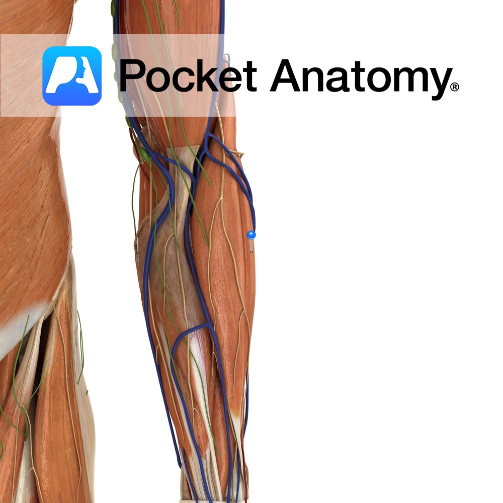
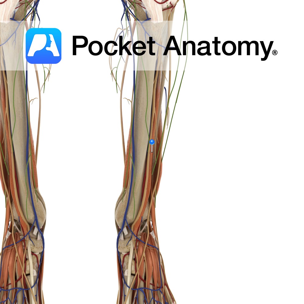
Anatomy Origin: Distal third of lateral supracondylar ridge of humerus and lateral intermuscular septum. Insertion: Posterior aspect of base of second metacarpal. Key Relations: One of the four muscles in the superficial posterior compartment of the forearm. Functions -Extends the hand at the wrist joint. -Abducts the hand at the wrist (radial deviation) particularly when
- Published in Pocket Anatomy Pins
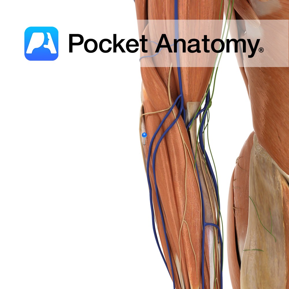
Anatomy Origin: Lateral epicondyle of humerus via common extensor tendon, adjacent intermuscular septa. Insertion: The dorsal aspect of base of second and third metacarpal. Key Relations: -One of the four muscles in the superficial posterior compartment of the forearm. Functions -Extends the hand at the wrist joint -Abducts the hand at the wrist (radial deviation)
- Published in Pocket Anatomy Pins

Anatomy Projects down from body of ethmoid (unpaired bone inside skull, between orbits, forming part cranial floor and part roof nasal cavity), forms part of nasal septum. Thin, flat, polygonal, usually deviated a little left or right. Septal cartilage attached below. Articulates with frontal, sphenoid, nasals. Clinical Ethmoid made up of perpendicular plate, cribriform plate
- Published in Pocket Anatomy Pins
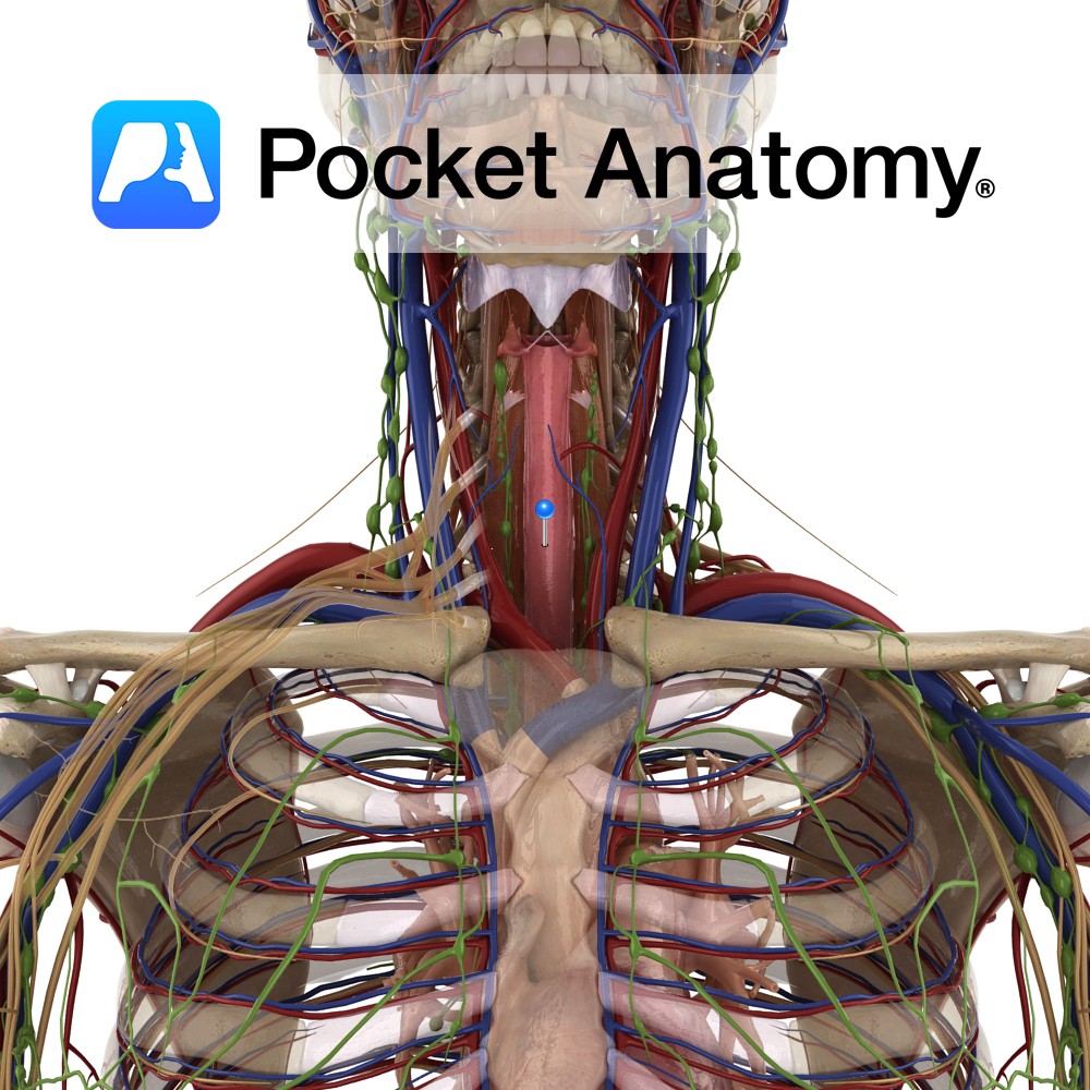
Anatomy Muscular tube, 4-12″, part of GI system (alimentary tract), connecting pharynx to stomach, continuous above with laryngeal pharynx at level C6; courses down behind trachea and heart, in front of spine, through mediastinum and on, passes through diaphragm at level T10, at which point it is continuous with stomach through a functional lower esophageal
- Published in Pocket Anatomy Pins
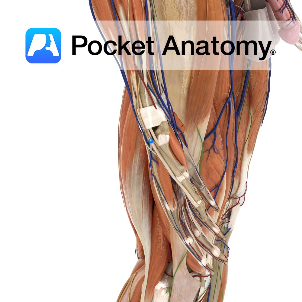
Anatomy Origin: Posterolateral aspect of middle third of ulna and adjacent interosseous membrane. Insertion: Dorsal surface of base of the distal phalanx of thumb. Key Relations: -The tendon of extensor pollicis longus passes underneath the extensor retinaculum in its own compartment. -Its tendon forms the posterior boundary of the anatomical snuffbox. -One of the six
- Published in Pocket Anatomy Pins
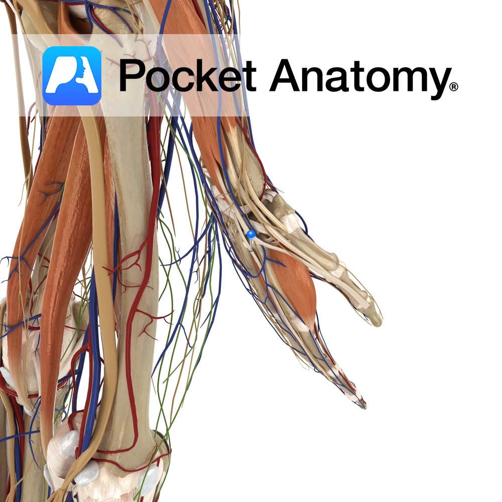
Anatomy Origin: Posterior of distal radius and adjacent Interosseus membrane. Insertion: Dorsal surface of base of proximal phalanx of thumb. Key Relations: -Its tendon together with the tendon of abductor pollicis longus forms the anterior boundary of the anatomical snuff box. -One of the six muscles in the deep posterior compartment of the forearm. Functions
- Published in Pocket Anatomy Pins
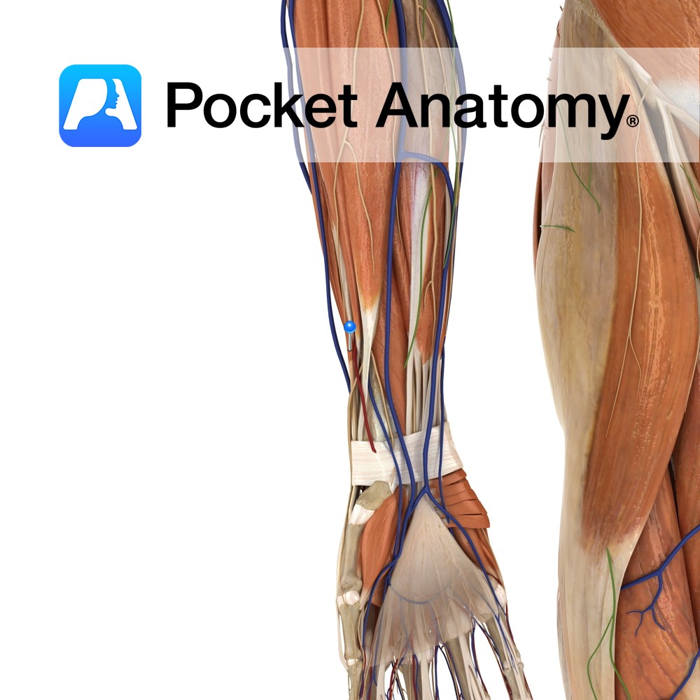
Anatomy Origin: Junction of middle and distal thirds of ulna and adjacent interosseous membrane. Insertion: Extensor hood of index finger. Key Relations: The tendon of extensor indicis passes under the extensor retinaculum in a compartment with extensor digitorum. One of the six muscles in the deep posterior compartment of the forearm. Functions -Extends index finger
- Published in Pocket Anatomy Pins
Anatomy Origin: Middle half of the anteromedial surface of the fibula and the interosseous membrane. Insertion: Dorsal surface of the base of the distal phalanx of the hallux (big toe). Key Relations: -One of the four muscles of the anterior compartment of the leg. -The extensor hallucis longus tendon crosses anterior to the ankle joint
- Published in Pocket Anatomy Pins
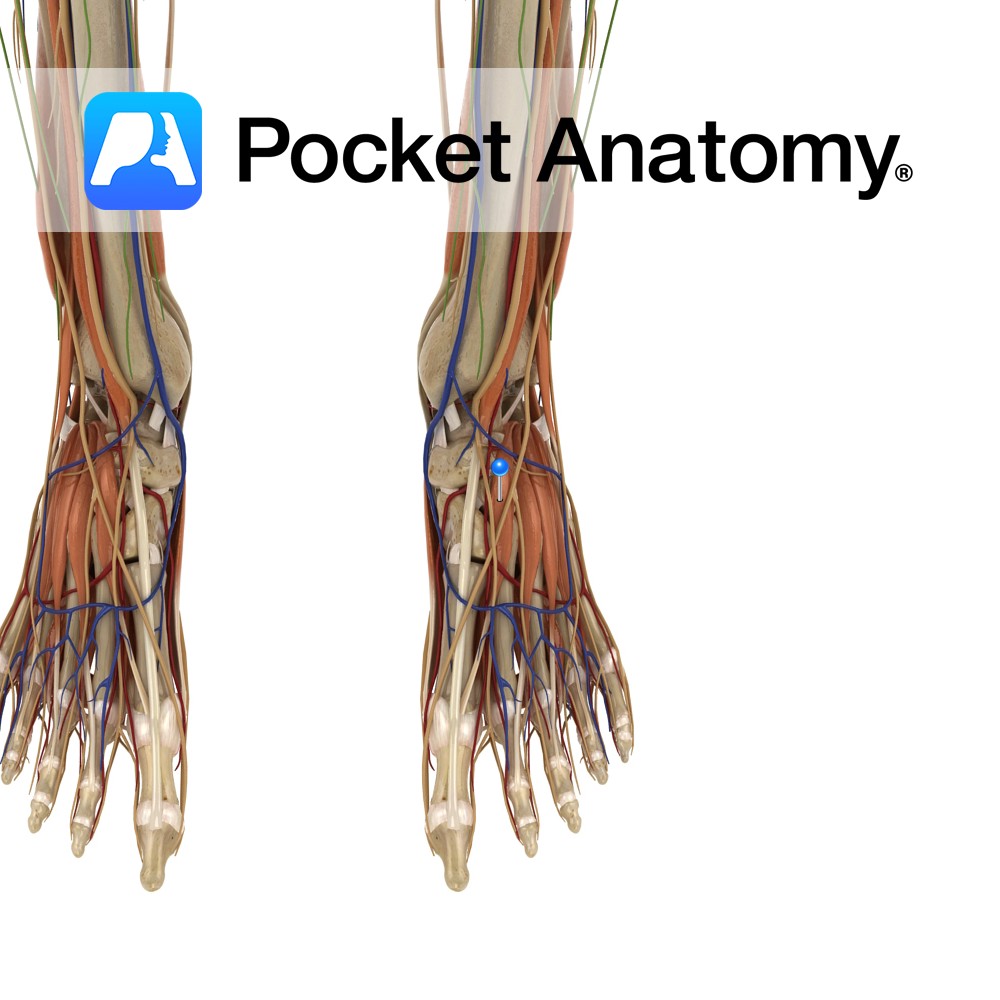
Anatomy Origin: Anterolateral part of the superior surface of calcaneus and inferior extensor retinaculum. Insertion: Dorsal surface of the base of the proximal phalanx of the hallux (big toe). Key Relations: Merely the medial part of extensor digitorum brevis. Functions Extends the hallux at the interphalangeal joint (assists extensor hallucis longus). Supply Nerve supply: Deep
- Published in Pocket Anatomy Pins