PocketAnatomy® is a registered brand name owned by © eMedia Interactive Ltd, 2009-2022.
iPhone, iPad, iPad Pro and Mac are trademarks of Apple Inc., registered in the U.S. and other countries. App Store is a service mark of Apple Inc.
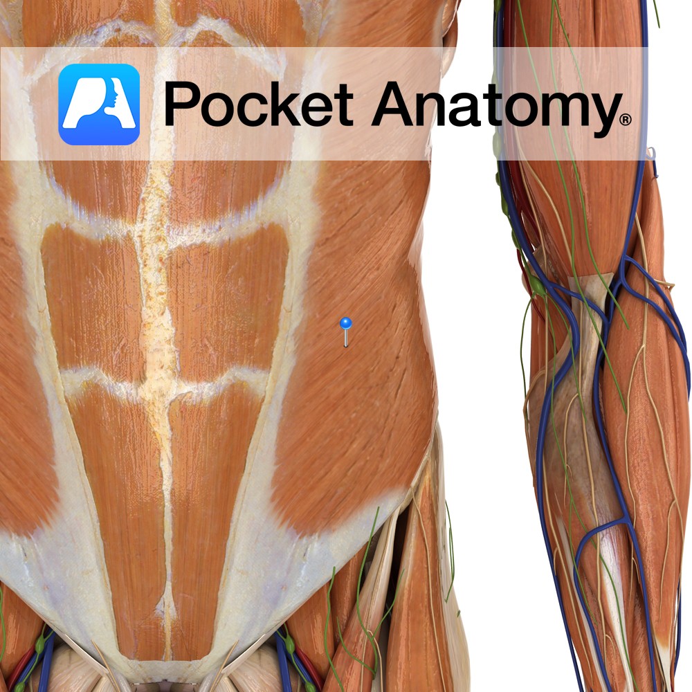
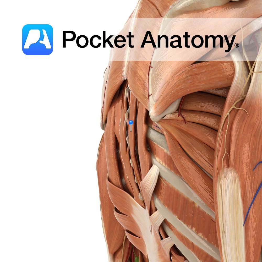
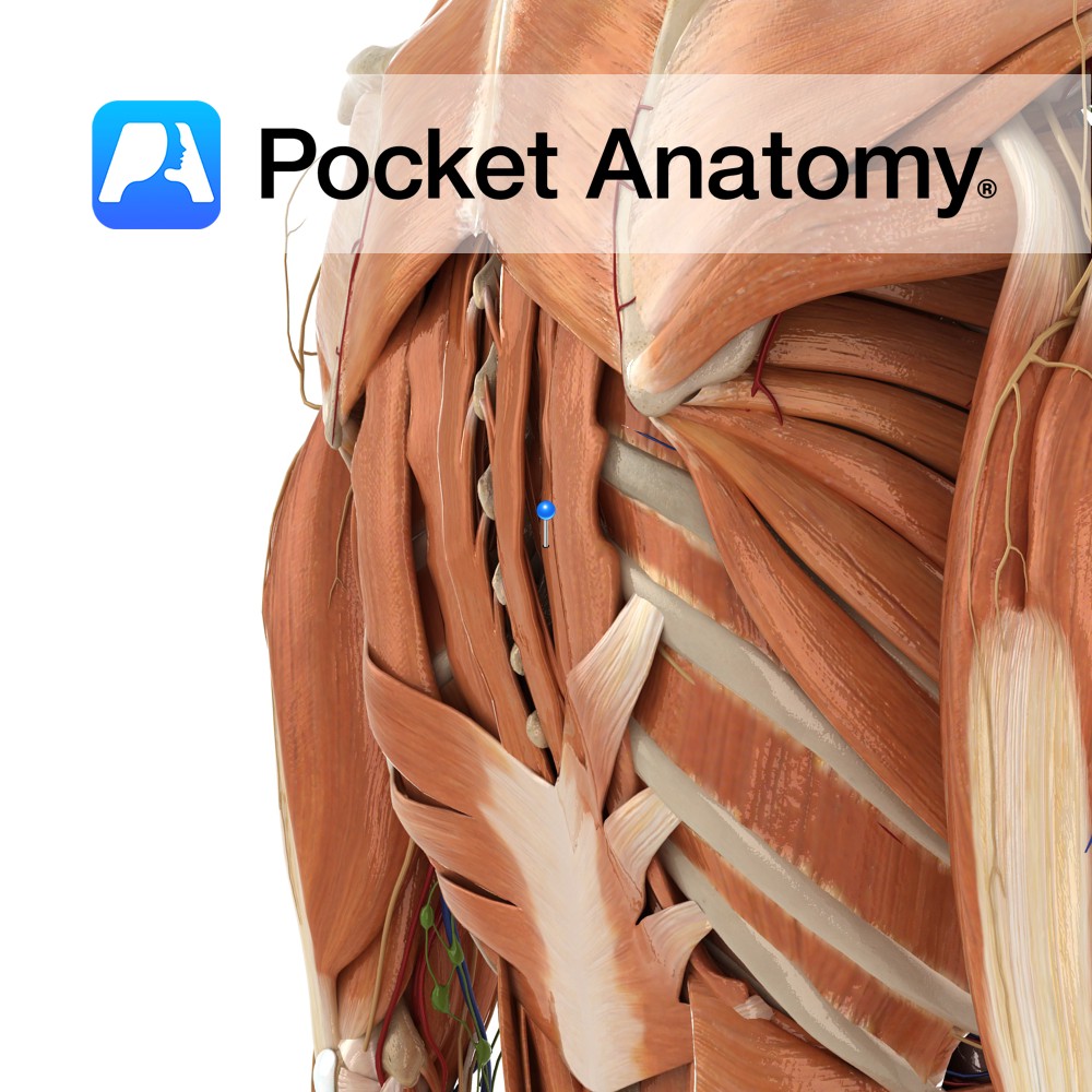
Anatomy Origin: Muscular digitations from the external inferior borders of the 5th to 12th ribs. Insertion: Via aponeurosis to the linea alba and the lateral lip of the iliac crest. Key relations: -Largest and most superficial of the three flat abdominal muscles. -Helpful to visualise the directions of the fibres as a putting your hands
- Published in Pocket Anatomy Pins
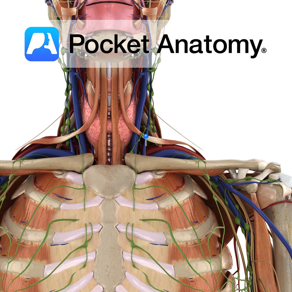
Anatomy Course Begins with the fusion of the posterior auricular vein and a portion of the retromandibular vein in the parotid gland. As it descends from the head it perforates the deep fascia until it eventually joins the subclavian vein, lateral to the internal jugular vein. Drain Drains the blood exterior to the cranium. Interested
- Published in Pocket Anatomy Pins
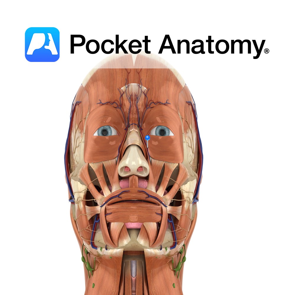
Anatomy Course Originates from the anterior side of the external carotid artery, where it then rises superiorly through the deep structures of the neck until it reaches the mandible. It then curves around the inferior border of mandible, anterior to the master muscle. From there it undergoes a tortuous path superomedially, where it becomes the
- Published in Pocket Anatomy Pins
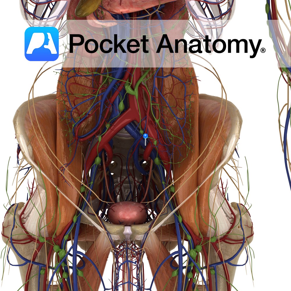
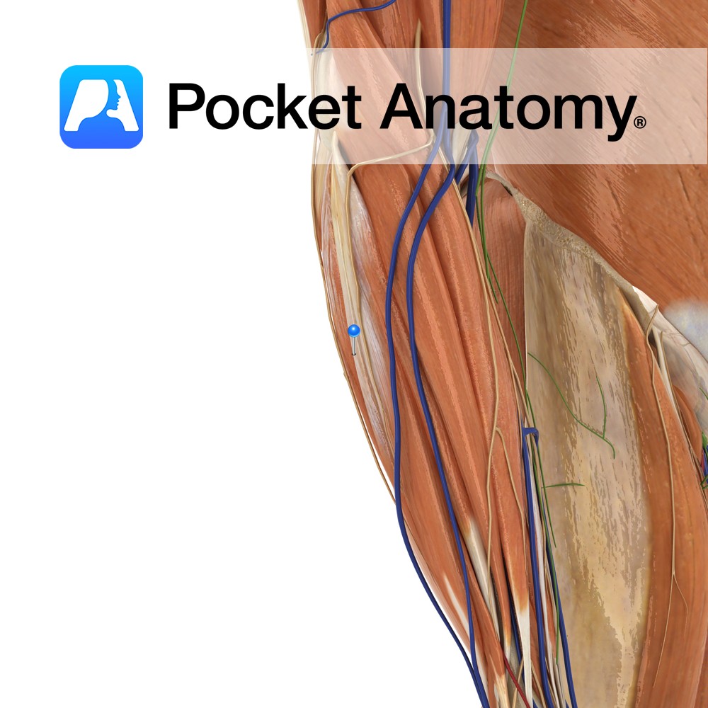
Anatomy Course Continuation of the femoral vein after it travels beneath the inguinal ligament. Travels briefly with its counterpart artery until it joins with the internal iliac vein and becomes the common iliac vein. Drain Drains the deep veins of the leg. Interested in taking our award-winning Pocket Anatomy app for a test drive?
- Published in Pocket Anatomy Pins
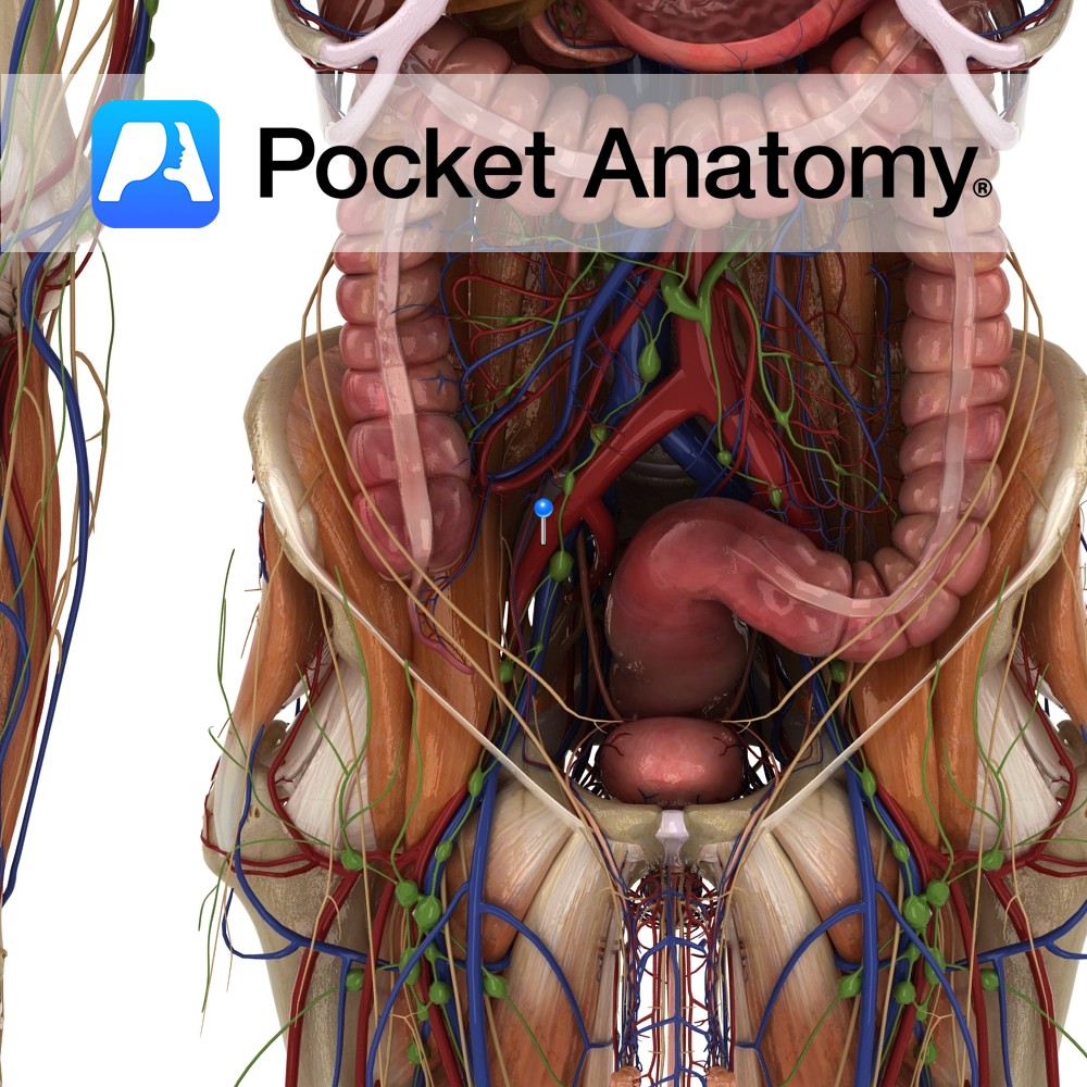
Anatomy Course Arises after the bifurcation of the common iliac artery. It descends on the psoas muscle until it passes underneath the inguinal ligament, where it is referred to as the femoral artery. Supply Major source for the lower limb. Clinical In renal transplants, the surgeon often attaches the renal artery of the donor’s kidney
- Published in Pocket Anatomy Pins

Anatomy The human eyes are the organs of vision, located in the bony orbits in the front of the skull, on either side of the bridge of the nose. Functions The eye detects light and transforms it into impulses that are sent to the brain. Light enters through an opening in the iris called the
- Published in Pocket Anatomy Pins
Anatomy One of the intrinsic muscles of the back. Consists of spinalis capitis, cervicus, thoracis. Spinalis capitis: Usually blends with semispinalis capitis. Spinalis cervicis: (Not always present) Origin: Ligamentum nuchae and spinous process of C7 (sometimes T1 to T2). Insertion: Spinous process of axis (C2). Spinalis thoracis: Origin: Spinous processes of L2 toT11. Insertion: Spinous
- Published in Pocket Anatomy Pins
Anatomy One of the intrinsic muscles of the back. Consists of longissimus capitis, cervicus and thoracis. Longissimus capitis: Origin: Transverse process of T1 to T5 and articular processes of C5 to C7 approx. Insertion: Posterior margin of mastoid process. Key Relations: Lies medial to longissimus cervicus. Longissimus cervicis: Origin: Transverse processes of T1 to T5.
- Published in Pocket Anatomy Pins
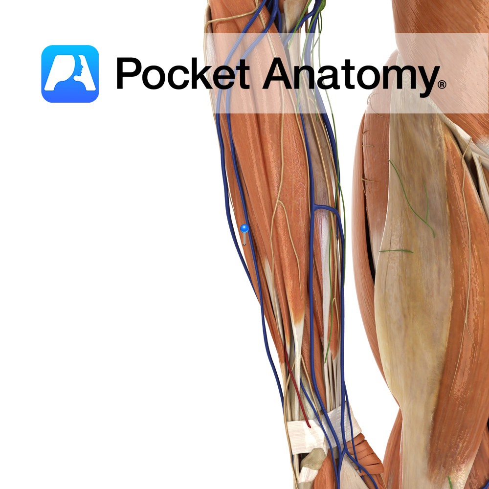
Anatomy Origin: Lateral epicondyle of humerus via the common extensor tendon and adjacent intermuscular septum. Insertion: Through separate tendons inserting into extensor hoods on dorsal surface of middle and distal phalanges of the four fingers. Key Relations: One of the two muscles in the intermediate posterior compartment of the forearm. Functions -Extension of four fingers
- Published in Pocket Anatomy Pins
Anatomy Origin: Lateral epicondyle of humerus via the common extensor tendon, adjacent intermuscular septum. Insertion: Dorsal hood of the little finger. Key Relations: One of the two muscles in the intermediate posterior compartment of the forearm. Functions -Extends the little finger at the metacarpophalangeal joint. -Extends the interphalangeal joints of the little finger. -Helps extend
- Published in Pocket Anatomy Pins