PocketAnatomy® is a registered brand name owned by © eMedia Interactive Ltd, 2009-2022.
iPhone, iPad, iPad Pro and Mac are trademarks of Apple Inc., registered in the U.S. and other countries. App Store is a service mark of Apple Inc.
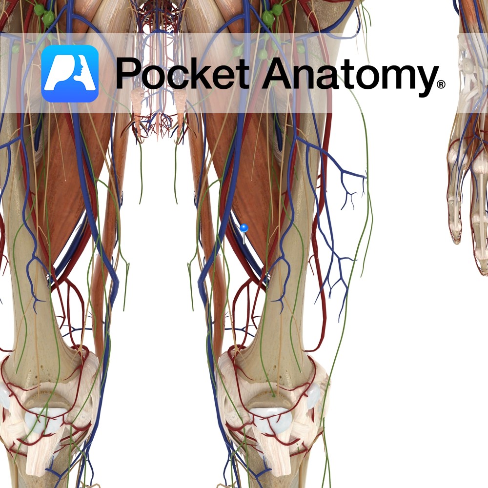
Anatomy Course Continuation of the popliteal artery when it enters the adductor canal. It travels along with its associated artery in the femoral sheath until it passes beneath the inguinal ligament, where it becomes the external iliac vein. Drain Drains the lower limb. Clinical Common site for injection of recreational drugs. Interested in taking our
- Published in Pocket Anatomy Pins
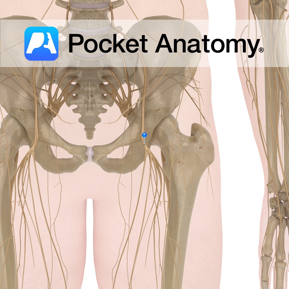
Anatomy Course The largest branch of the lumbar plexus and is composed of the second, third and fourth lumbar nerves. Descends between the iliacus muscle and psoas major muscle and runs below the inguinal ligament where it divides into two branches, anterior and posterior. Supply Motor supply to the quadriceps femoris as well as many
- Published in Pocket Anatomy Pins
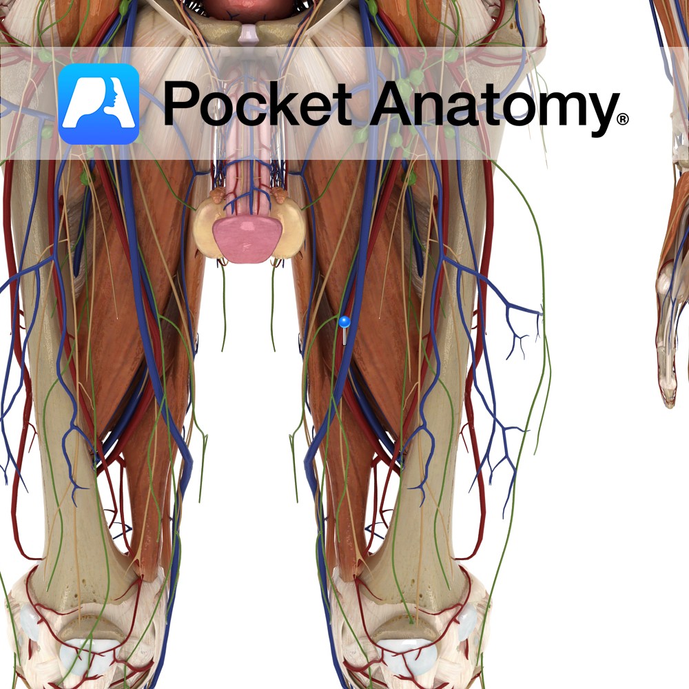
Anatomy Course The external iliac artery becomes the femoral artery as it passes below the inguinal ligament and enters the femoral triangle, where it then descends in the adductor canal. It eventually exits via the adductor hiatus of the adductor magnus muscle, where it becomes the popliteal artery. Supply Supplies cutaneous regions of the lower
- Published in Pocket Anatomy Pins
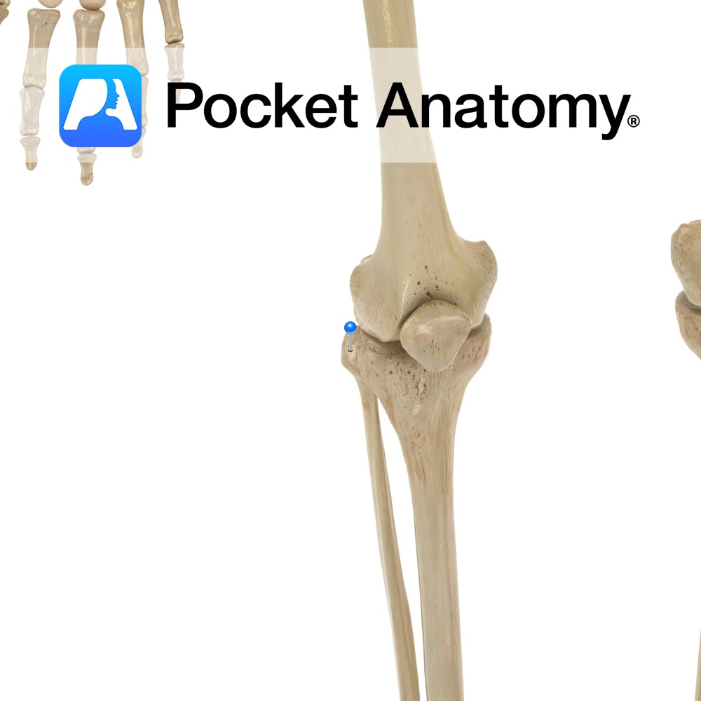
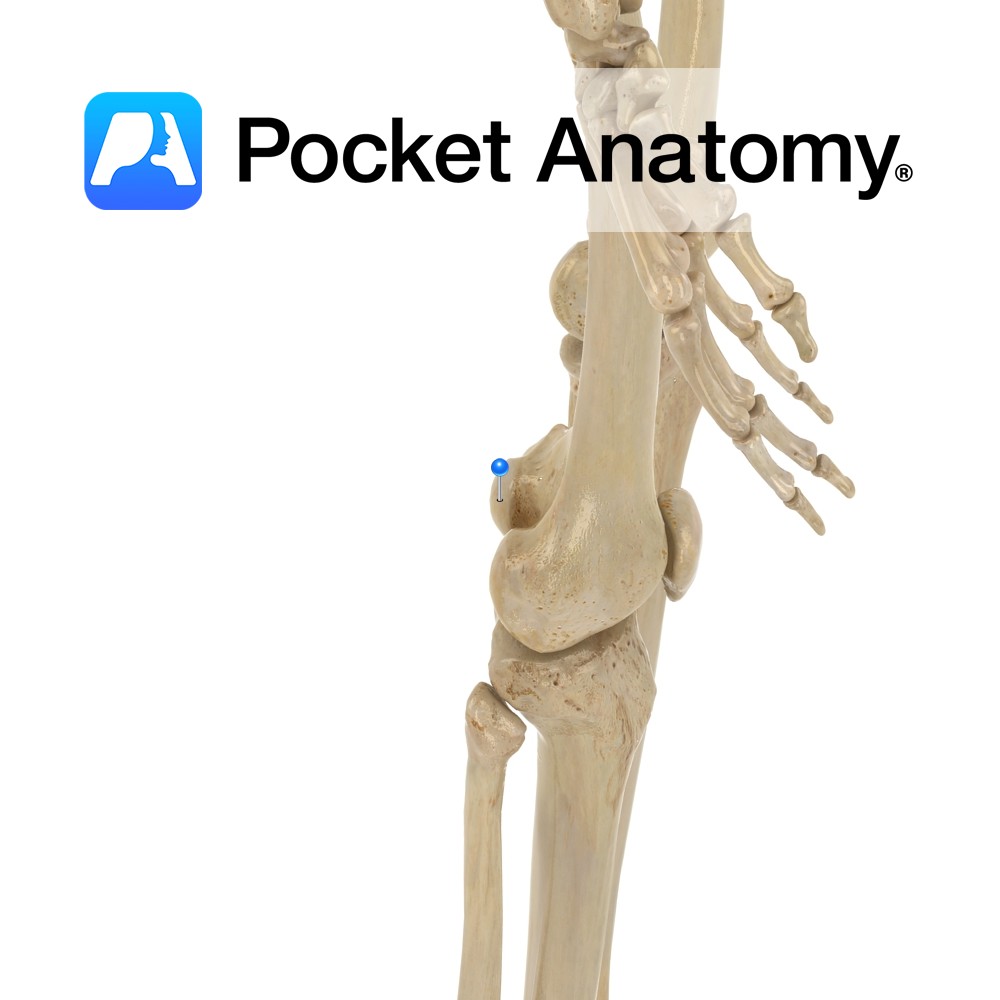
Anatomy Small, quadrate, lower and more posterior than lateral condyle tibia. Connected to lateral condyles of femur (by fibular – also known as lateral – collateral ligament) and tibia (proximal tibiofibular joint and anterior and posterior ligaments). Clinical Lateral collateral ligament (LCL) injury less common than medial, as force required on inside of knee (to
- Published in Pocket Anatomy Pins
Anatomy Cylindrical, courses down and in (hips are further apart than knees, more so in female). Clinical Shaft is very strong, surrounded by large richly perfused muscles. Takes a lot of force to fracture (spiral, comminuted or open). Displacement of fragments usual (muscles pull); surgery required. Life-threatening blood loss can occur in open fracture; significant
- Published in Pocket Anatomy Pins
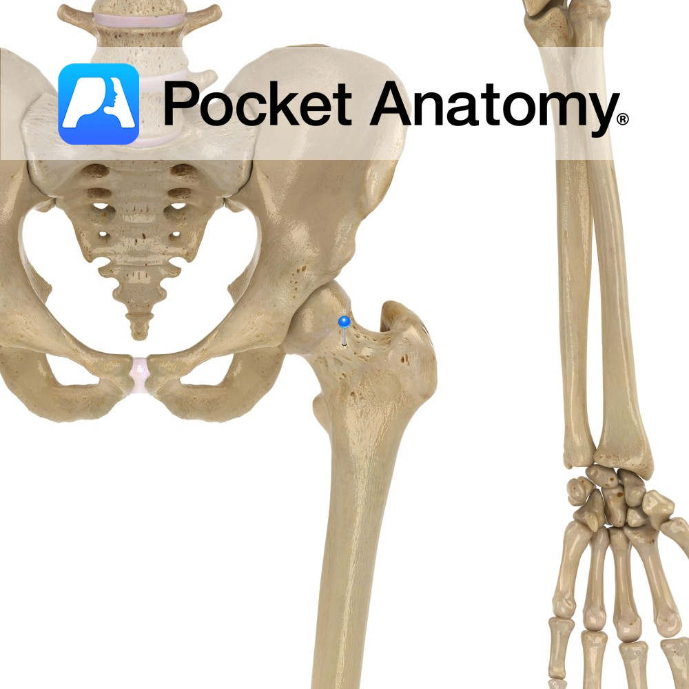
Anatomy Angle of 125° with shaft in adulthood; angle less in shorter femurs and wider pelvises (sa female). Points up, in and forward (12 – 14°). Big load-bearing and torsion associated with locomotion, are withstood by relatively small neck bone mass, through its tension and compression trabeculae and strong calcar femorale. Clinical Classicly presents as:
- Published in Pocket Anatomy Pins
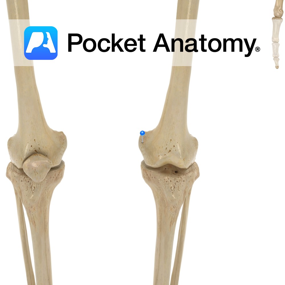
Anatomy Convex bump at inner/medial aspect of lower femur (bigger, more prominent than lateral epicondyle). Attachments; tibial (medial) collateral ligament (MCL), which connects to lateral condyle tibia, medial head of gastrocnemius behind. Interested in taking our award-winning Pocket Anatomy app for a test drive?
- Published in Pocket Anatomy Pins
Anatomy Oblong eminence at medial/inner aspect of lower femur. Separated from lateral condyle in front by patellar space (trochlear groove), at back by intercondyloid fossa. Articulates at front with patella, bottom and back with tibia. Clinical Cartilage tears (osteochondral fractures) can occur in particular in the knee (medial condyle more than lateral, often as part
- Published in Pocket Anatomy Pins
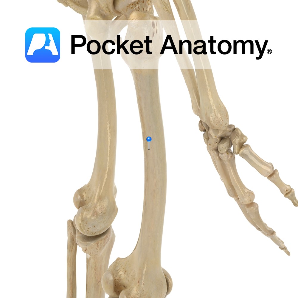
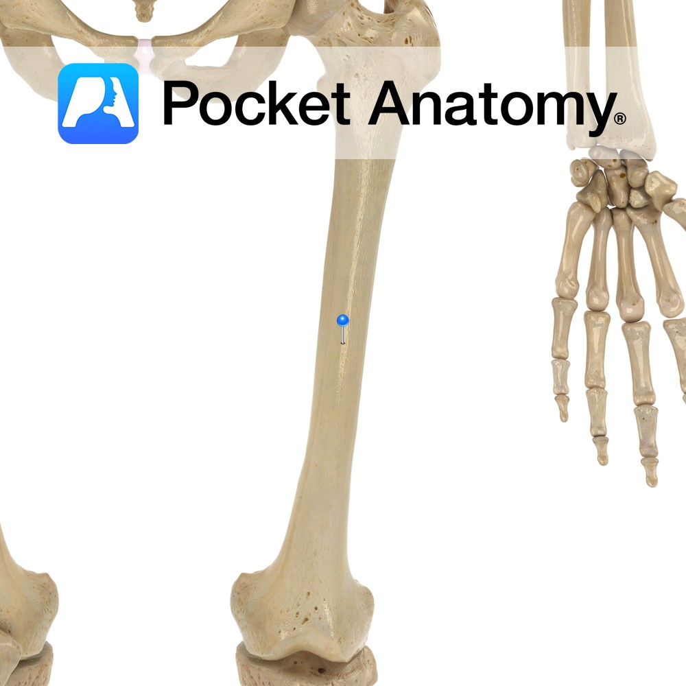
Anatomy Longitudinal ridge/crest at back of shaft. Many muscles attach: Medial lip and its extensions up and down – vastus medialis; lateral lip and its extension up – vastus lateralis; intermediate line, lateral extension above and medial extension below – adductor magnus; between vastus lateralis and adductor magnus – gluteus maximus above, short head biceps
- Published in Pocket Anatomy Pins
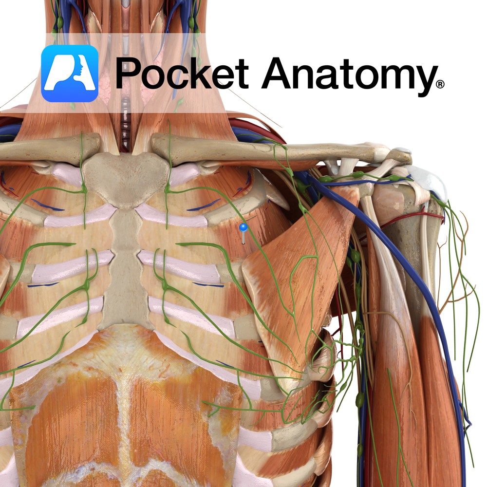
Anatomy Origin: Inferior margin of the rib above i.e. ribs 1 to 11. Insertion: Superior surface of the rib below i.e. ribs 2 to 12. Key relations: -Muscle fibres run obliquely anteroinferiorly. -Most superficial intercostal muscle. Functions -Moves ribs superiorly during inspiration, thereby expanding the thoracic cavity. -Maintains intercostals spaces during inspiration and expiration. Supply
- Published in Pocket Anatomy Pins