PocketAnatomy® is a registered brand name owned by © eMedia Interactive Ltd, 2009-2022.
iPhone, iPad, iPad Pro and Mac are trademarks of Apple Inc., registered in the U.S. and other countries. App Store is a service mark of Apple Inc.
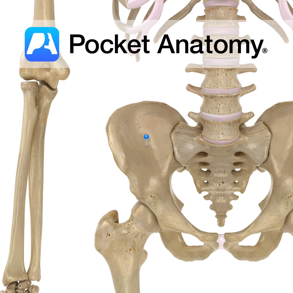
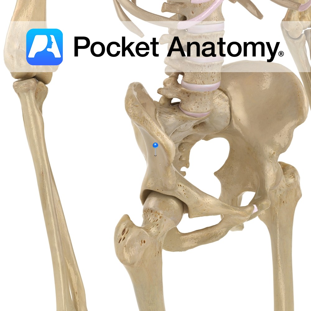
Anatomy The ala (wing) of the ilium is curved, forming a large concave fossa on its internal surface, its boundaries being the iliac crest, arcuate line and anterior and posterior borders. Interested in taking our award-winning Pocket Anatomy app for a test drive?
- Published in Pocket Anatomy Pins
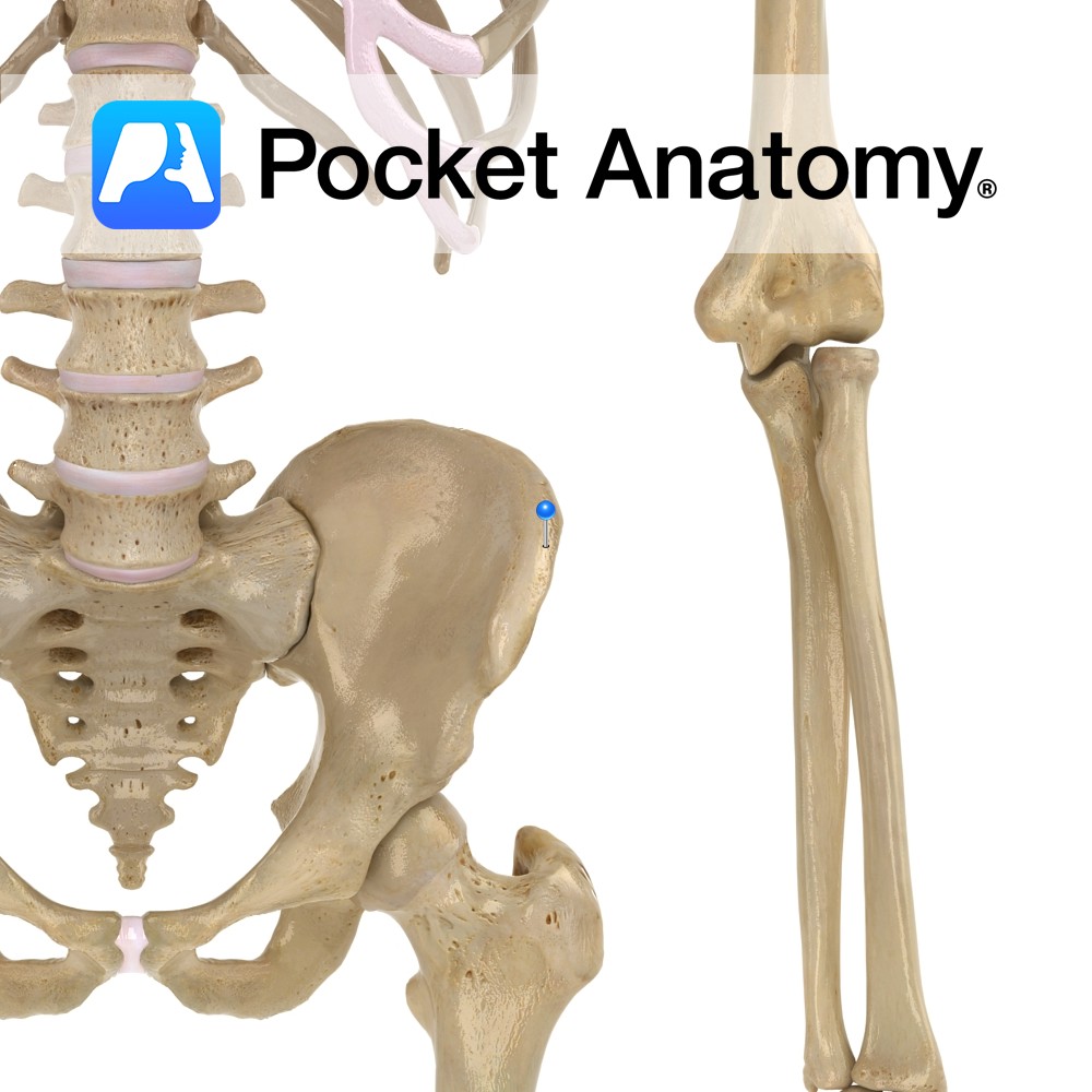
Anatomy The top (superior border) of the ilium, convex front to back (from anterior superior iliac spine to posterior superior iliac spine), sinuous side-to-side (concave in at front, concave out behind). Clinical The crest is palpable throughout its length; multiple muscles and fascia attach to its inner lip, intermediate line and outer lip. Vignette Standing
- Published in Pocket Anatomy Pins
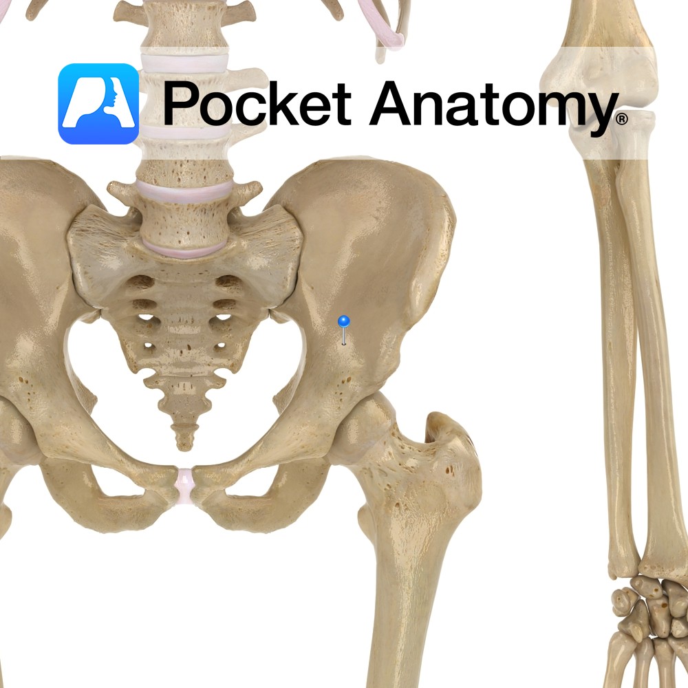
Anatomy The outside (lateral) surface of the wing (ala) of the ilium, site for attachment (from back to front) of gluteus maximus, medius, minimus. Interested in taking our award-winning Pocket Anatomy app for a test drive?
- Published in Pocket Anatomy Pins
Anatomy The pelvic brim (edge of pelvic inlet) is formed by the arcuate lines of the ilia, the pectineal lines of the pubis, the upper margin of the pubic symphysis and the sacral promontory. Above, abdominal cavity; below, pelvic cavity. Vignette Viewed from above, the pelvic brim is smooth, almost circular. Interested in taking our
- Published in Pocket Anatomy Pins
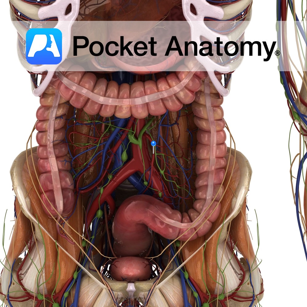
Anatomy Course Branches off the anterior aspect of the abdominal aorta at the level of L3. It briefly descends on the abdominal aorta before crossing to the left and giving off its branches. Supply Supplies a portion of the colon that commences at the splenic flexure to the upper rectum. Interested in taking our award-winning
- Published in Pocket Anatomy Pins
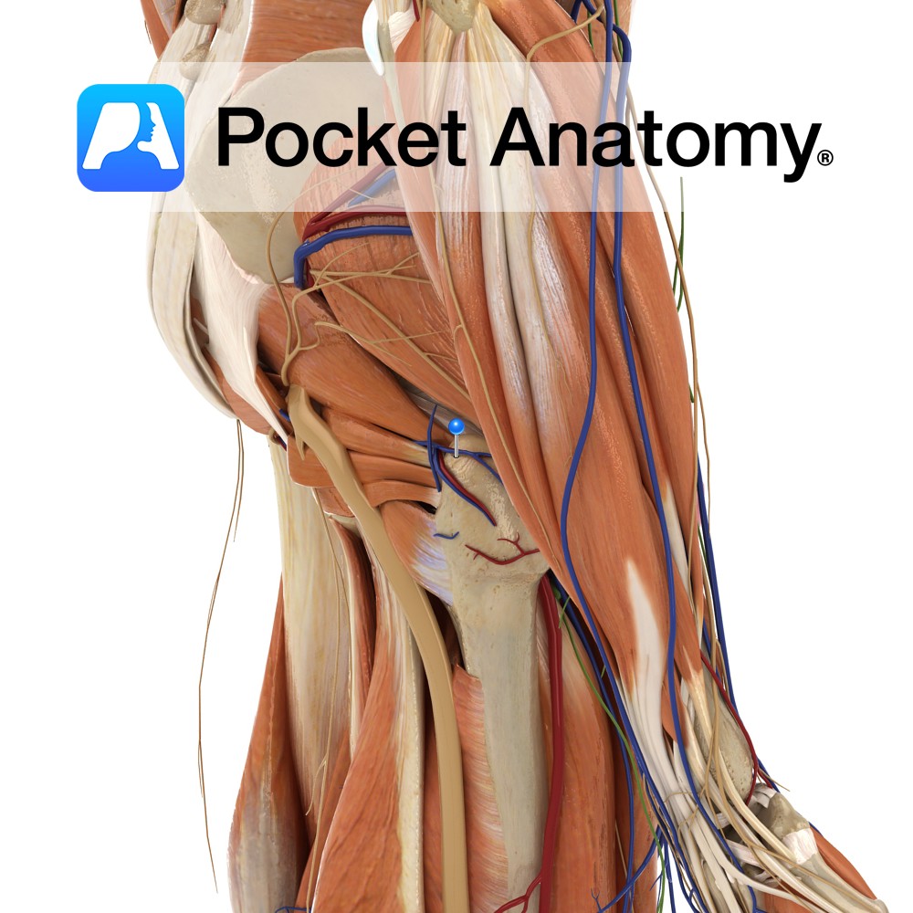
Anatomy Course Originates on the superior posterior surface of the thigh where they create an anastomotic network with the medial femoral circumflex vein. To enter the pelvis they pass through the greater sciatic foramen below the piriformis muscles and empty into the hypogastric vein. Drain Drains the inferior aspect of the gluteal region. Interested in
- Published in Pocket Anatomy Pins
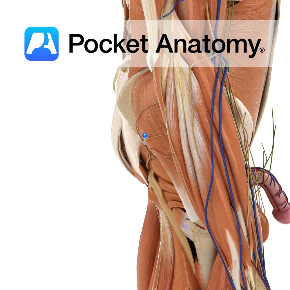
Anatomy Course Originates from the sacral plexus, from fibres that originate from the fifth lumbar and first and second sacral nerves. From there it exits the pelvis through the greater sciatic foramen superior to the piriformis. Supply Innervates the gluteus maximus and deep gluteal muscles. Interested in taking our award-winning Pocket Anatomy app for a
- Published in Pocket Anatomy Pins
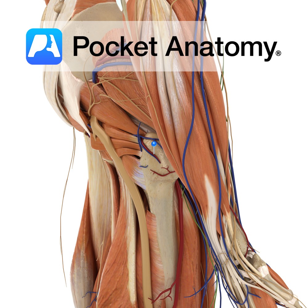
Anatomy Course One of the terminal branches of the internal iliac artery. Exits the pelvic cavity via the greater sciatic foramen, below the piriformis muscle, to enter the gluteal region. Supply Mainly supplies the gluteal region and the posterior compartment of the thigh. Also supplies the sciatic nerve. Interested in taking our award-winning Pocket Anatomy
- Published in Pocket Anatomy Pins
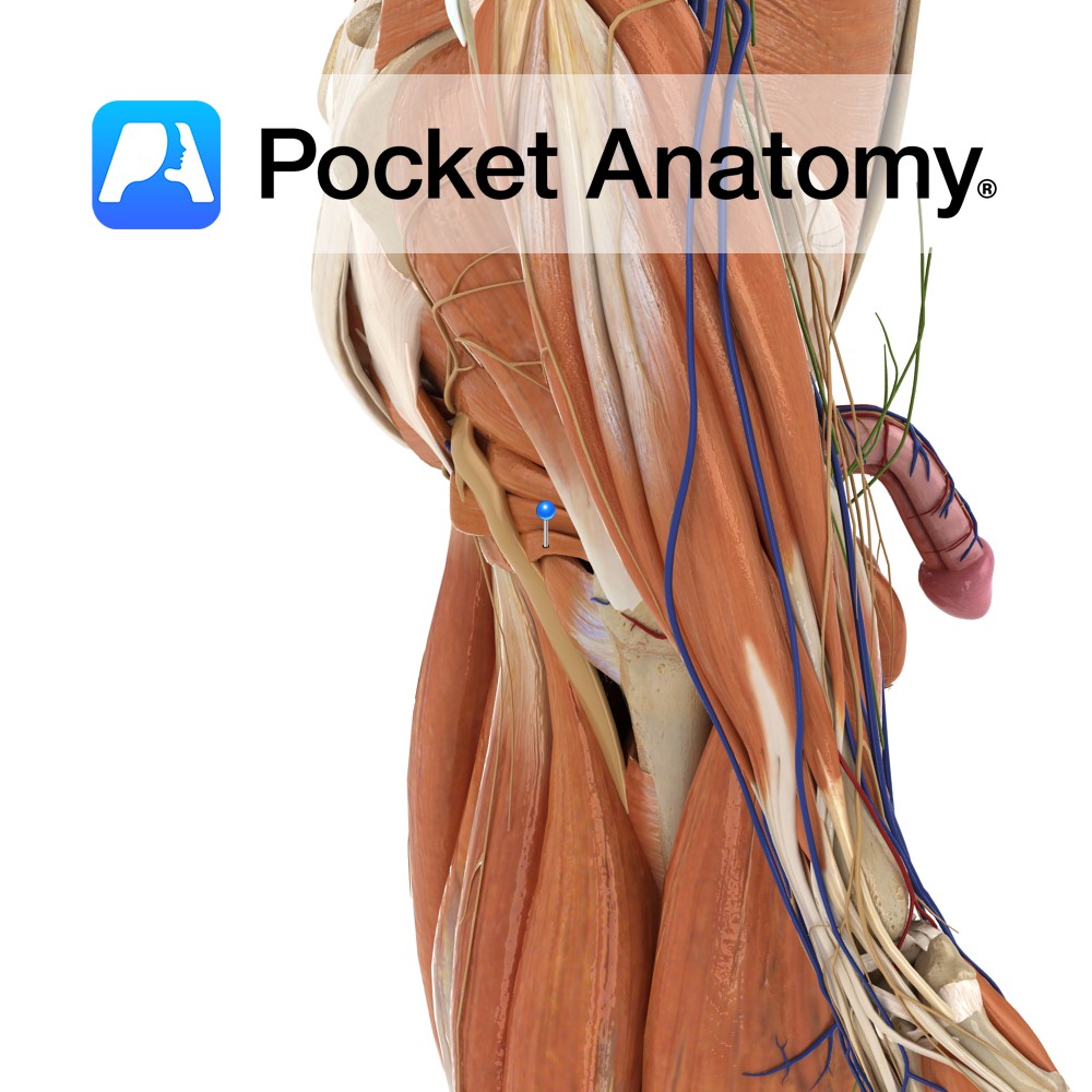
Anatomy Origin: Upper part of the ischial tuberosity, immediately below the groove for the obturator internus tendon. Insertion: Blends with the more posterior fibres of the tendon of the obturator internus and attaches with the tendon to the medial surface of the greater trochanter of the femur. Key Relations: Lies inferior to the obturator internus
- Published in Pocket Anatomy Pins
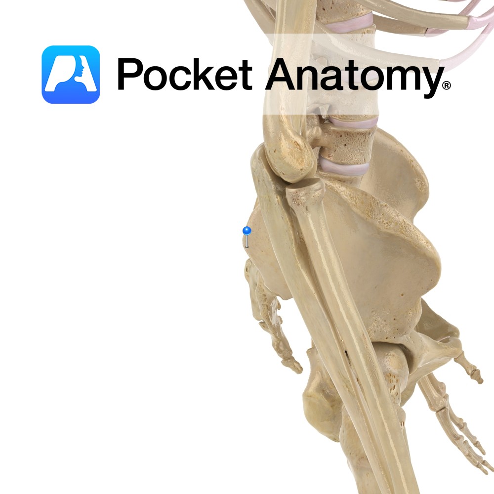
Anatomy Bulge on the back border of the crest of the ala of the ilium, separated by a notch below from the posterior inferior iliac spine. Interested in taking our award-winning Pocket Anatomy app for a test drive?
- Published in Pocket Anatomy Pins

