PocketAnatomy® is a registered brand name owned by © eMedia Interactive Ltd, 2009-2022.
iPhone, iPad, iPad Pro and Mac are trademarks of Apple Inc., registered in the U.S. and other countries. App Store is a service mark of Apple Inc.
.jpg)
Anatomy Articulates up with 3rd proximal and down with 3rd distal phalanges. Interested in taking our award-winning Pocket Anatomy app for a test drive?
- Published in Pocket Anatomy Pins
.jpg)
Anatomy Articulates posteriorly with 5th proximal and anteriorly with 5th distal phalanges. Interested in taking our award-winning Pocket Anatomy app for a test drive?
- Published in Pocket Anatomy Pins
.jpg)
Anatomy Articulates up/proximally with proximal phalanx at 3rd PIP, and distally with distal phalanx at 3rd distal interphalangeal joint (DIP). Interested in taking our award-winning Pocket Anatomy app for a test drive?
- Published in Pocket Anatomy Pins
.jpg)
Anatomy Articulates up/proximally with proximal phalanx at 5th PIP, and distally with distal phalanx at 5th distal interphalangeal joint (DIP). Interested in taking our award-winning Pocket Anatomy app for a test drive?
- Published in Pocket Anatomy Pins
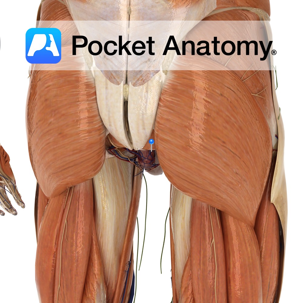
Anatomy Course A branch of the anterior trunk of the internal iliac artery. It branches from the anterior trunk before the anterior trunk passes between the rami anterior rami of two of the sacral nerves. It courses medially along the posterior pelvic wall towards the rectum, where it joins with the superior and inferior rectal
- Published in Pocket Anatomy Pins
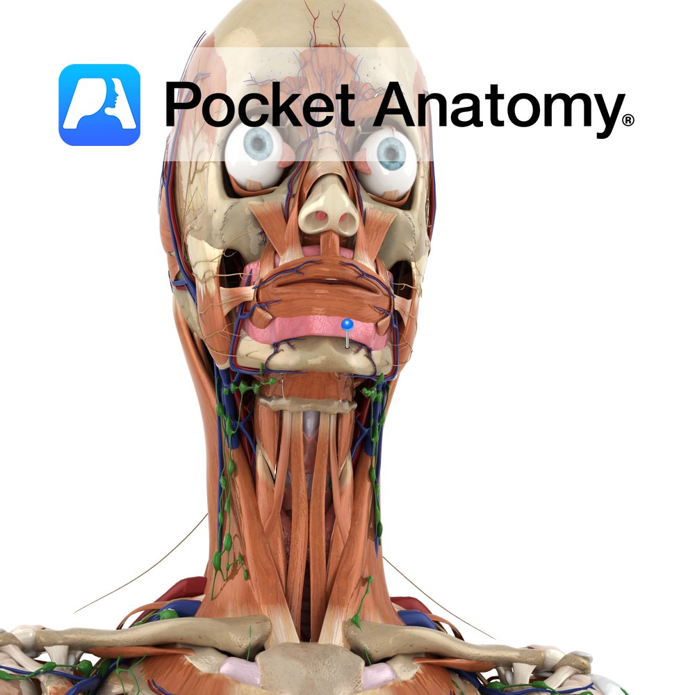
Anatomy Course A continuation of the inferior alveolar nerve, the mental nerve exits the mental foramen of the mandible. Supply Provides sensation to the anterior aspects of the chin, lower lip, molar and premolar teeth as well as related gingiva. Interested in taking our award-winning Pocket Anatomy app for a test drive?
- Published in Pocket Anatomy Pins
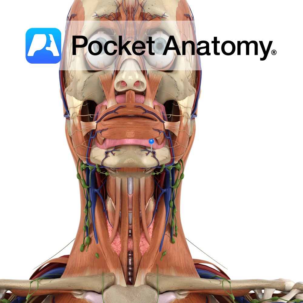
Anatomy Course Branch of the inferior alveolar vein which travels with the mental nerve and mental artery through the mental foramen of the mandible. Drain Responsible for the drainage of the lower teeth and jaw. Interested in taking our award-winning Pocket Anatomy app for a test drive?
- Published in Pocket Anatomy Pins
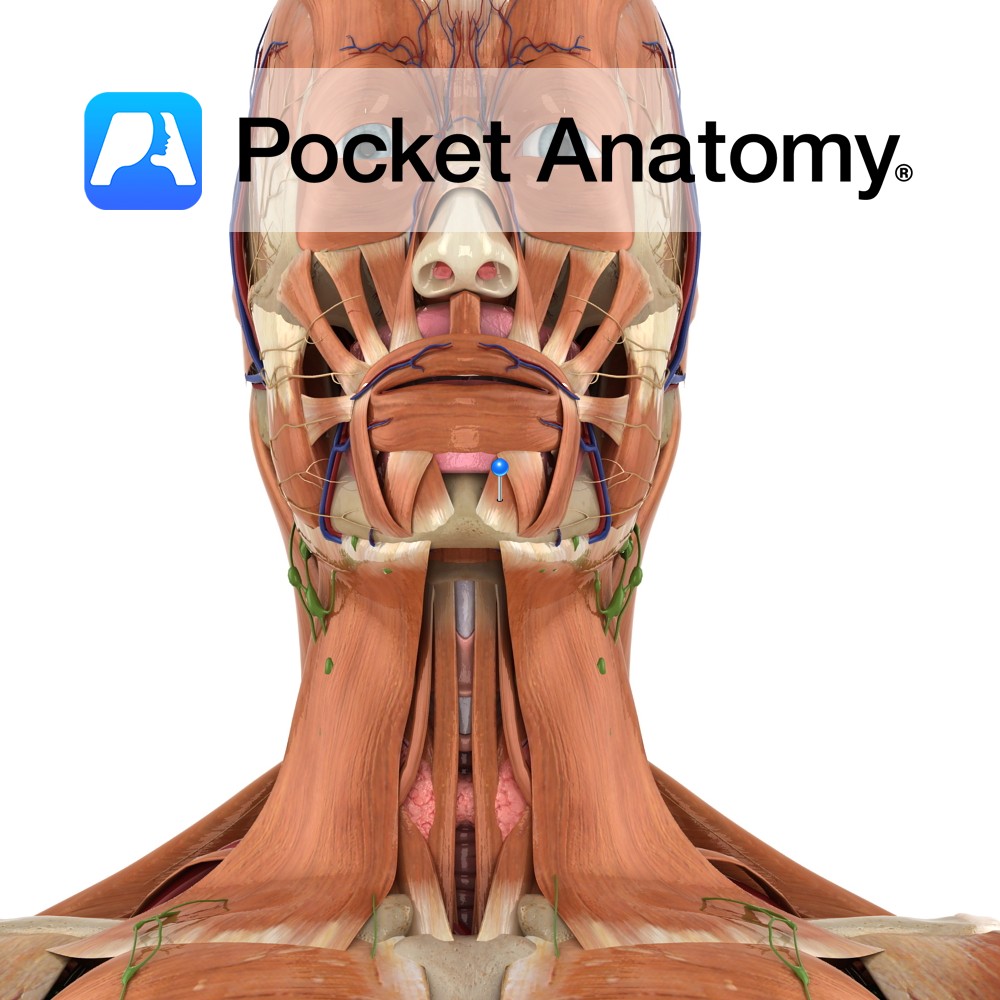
Anatomy Origin: Mandible inferior to the incisor teeth. Insertion: Skin of the chin. Key Relations: Deepest muscle along the mandible. Functions Raises and protrudes the lower lip as it wrinkles the skin of chin. e.g. when pouting or drinking from a glass.. Supply Nerve Supply: Mandibular branch of the facial nerve (CN 7). Blood Supply:
- Published in Pocket Anatomy Pins
.jpg)
Anatomy At near end is 1st (thumb) carpometacarpal joint (with trapezium), a saddle joint, allowing biggest range of finger movement, including opposition. At far end is 1st metacarpophalangeal joint (MCP). Interested in taking our award-winning Pocket Anatomy app for a test drive?
- Published in Pocket Anatomy Pins
.jpg)
Anatomy Longest metacarpal. Articulates; up (proximally) with (out to in, ie laterally to medially) trapezium, trapezoid, capitate: in (just below base) with 3rd metacarpal: down (distally) with 2nd proximal phalanx. Clinical 2nd metacarpal susceptible to injury from direct trauma, such as a hockey stick slash, or cricket ball on batsman’s hand. Interested in taking our
- Published in Pocket Anatomy Pins