PocketAnatomy® is a registered brand name owned by © eMedia Interactive Ltd, 2009-2022.
iPhone, iPad, iPad Pro and Mac are trademarks of Apple Inc., registered in the U.S. and other countries. App Store is a service mark of Apple Inc.
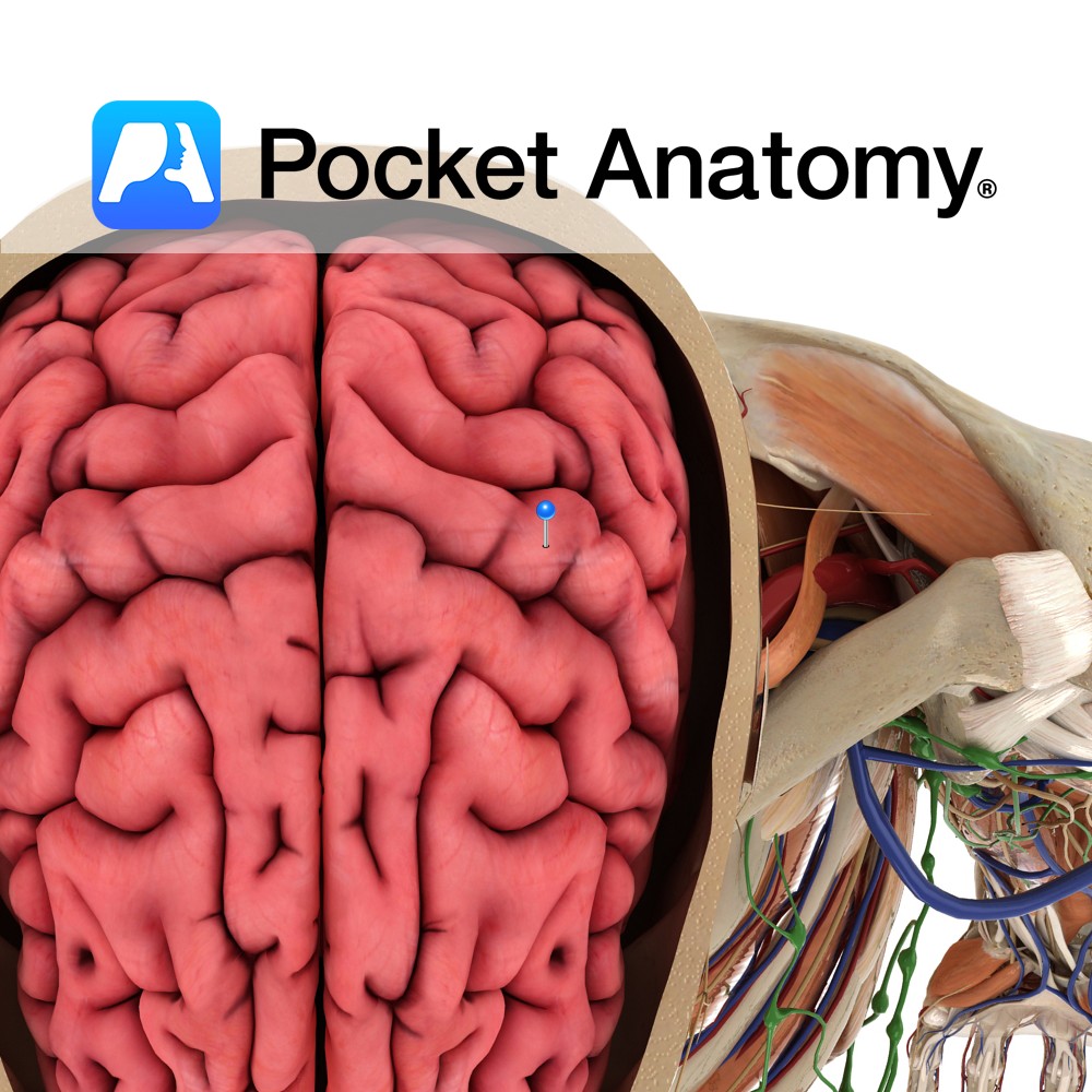
Anatomy A gyrus located posterior to the central sulcus. It extends anteroinferiorly from the great longitudinal fissure and ends just above the lateral sulcus. It forms part of the parietal lobe. Blood supply: Supplied by the middle cerebral artery, and medially by the anterior cerebral artery, to a lesser extent. Functions Is the location of
- Published in Pocket Anatomy Pins
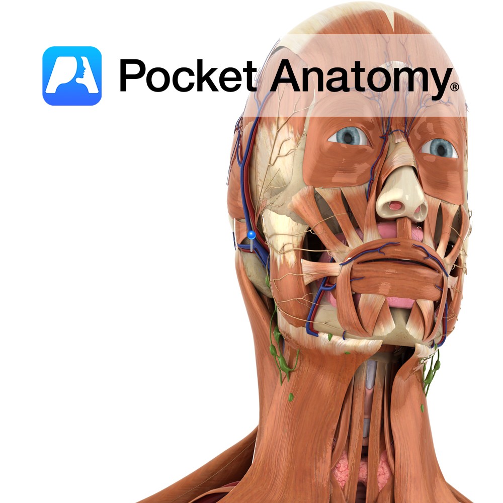
Anatomy Course A branch of the external carotid artery. It branches from the external carotid just above the posterior belly of the digastric muscle. It then ascends posteriorly behind the parotid gland, near the cartilage of the ear. Supply Supplies the auricle of the ear, as well as some of the scalp behind it. Interested
- Published in Pocket Anatomy Pins
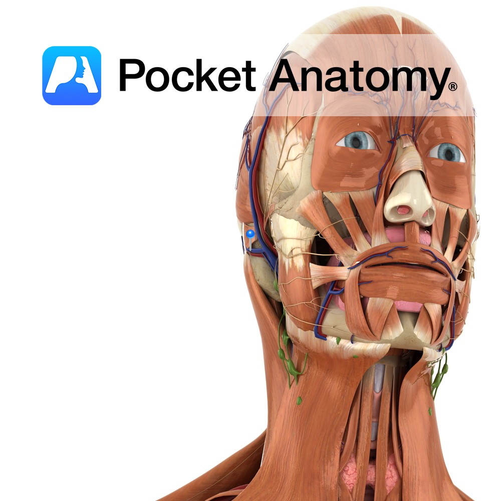
Anatomy Course Begins from a plexus on the side of the scalp. It descends behind the ear to join with the posterior facial vein. These both form the external jugular vein. Drain Drains the blood from the region of the scalp behind the ear. Interested in taking our award-winning Pocket Anatomy app for a test
- Published in Pocket Anatomy Pins
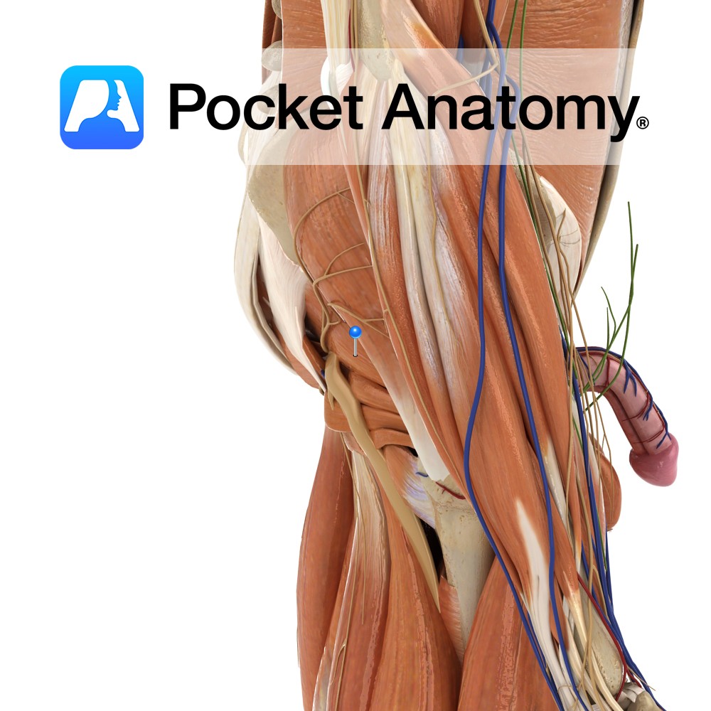
Anatomy Origin: Anteriolateral surface of the sacrum by three digitations, greater sciatic notch and sacrotuberous ligament. Insertion: After passing through the greater sciatic foramen, inserts into medial side on the superior border of the greater trochanter of the femur. Key Relations: -Divides the greater sciatic foramen. Only the superior gluteal nerve passes superior to it.
- Published in Pocket Anatomy Pins
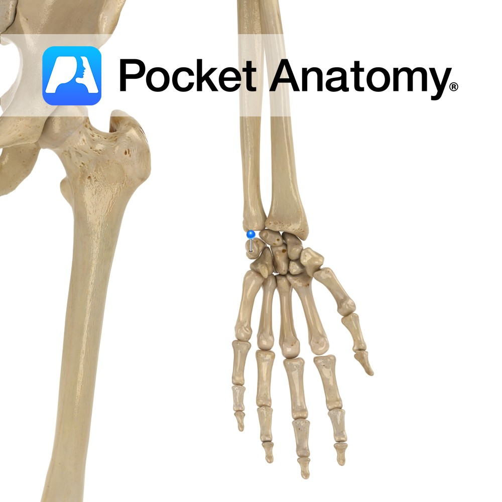
Anatomy Small finger side (ulnar) of nearer (proximal) row of carpal bones, pea-shaped. Articulates up and in with triquetral. Attachments; transverse carpal ligament, flexor carpi ulnaris, abductor digiti quinti. Sesamoid bone (a bone inside a tendon that passes over a joint, protecting it and providing effective load transmission) in ulnar collateral ligament. Clinical The wrist
- Published in Pocket Anatomy Pins
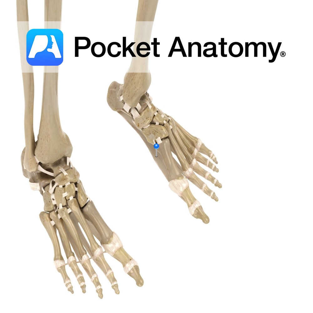
Anatomy Fascia consisting of thick connective tissue, of which most are longitudinally oriented collagen fibers. Attaches posteriorly to the tuberosity of the calcaneus, and anteriorly to the metatarsals. The fascia has three components: medial, central (which is the largest), and lateral. Functions Supports plantar aspect of the foot, and helps keep the arch of the
- Published in Pocket Anatomy Pins
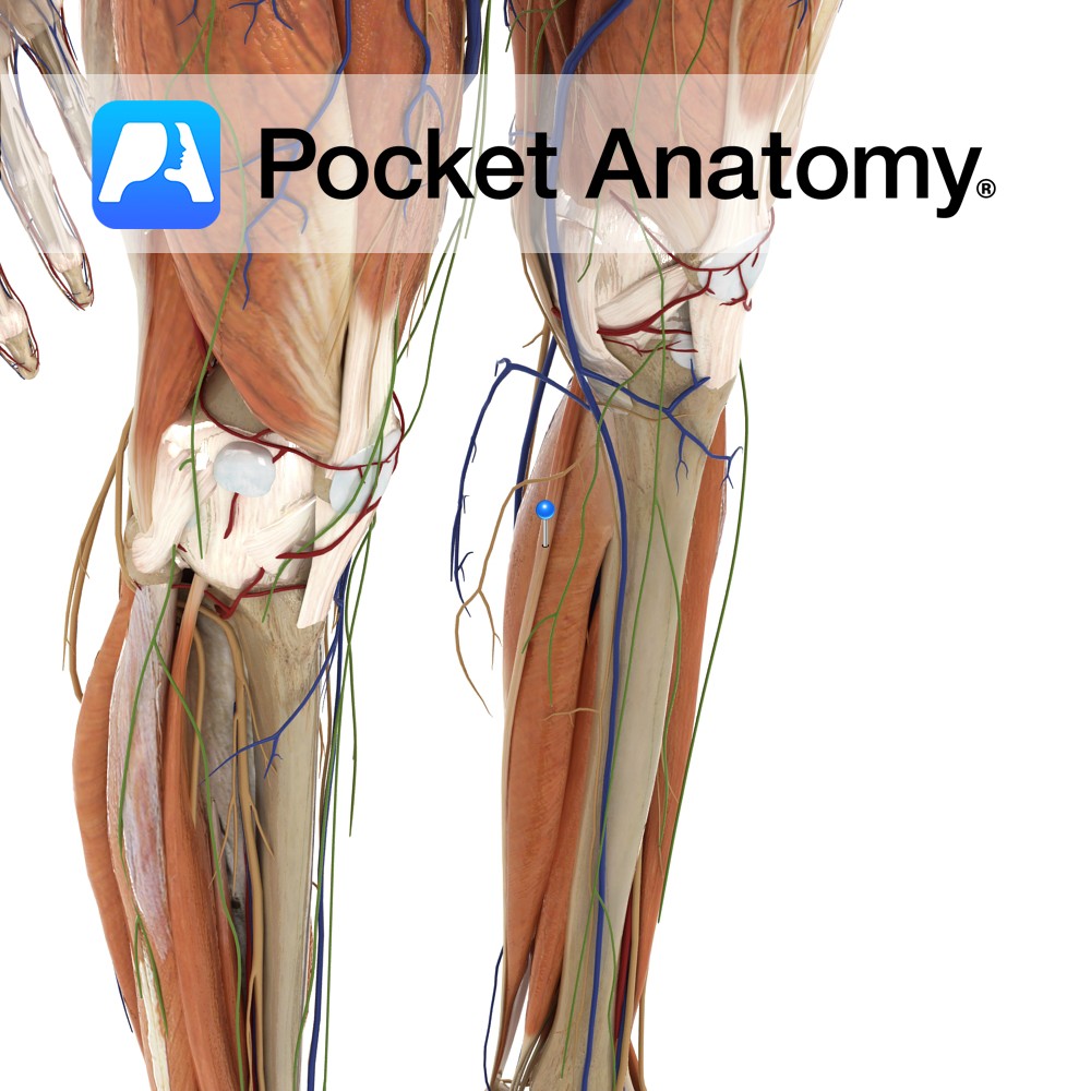
Anatomy Origin: Inferior aspect of the lateral supracondylar line of the femur and the popliteal surface of the femur. Insertion: Medial side of posterior surface of calcaneus by the tendo calcaneus (Achilles tendon). Key Relations: -One of the three muscles of the superficial posterior compartment of the leg. -Forms the lateral lower border of the
- Published in Pocket Anatomy Pins
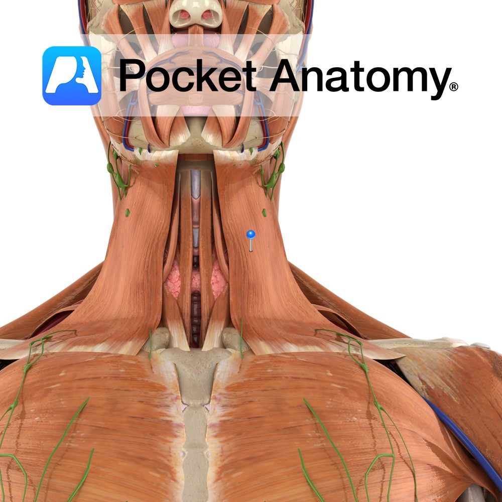
Anatomy Origin: Superficial fascia of the upper part of the thorax, covering pectoralis minor and deltoid. Insertion: Lower border of the body of the mandible, skin of the lower face, lateral half of the lower lip, and the buccal angle. Key Relations: -Platysma runs from the root of the neck across the clavicle to the
- Published in Pocket Anatomy Pins
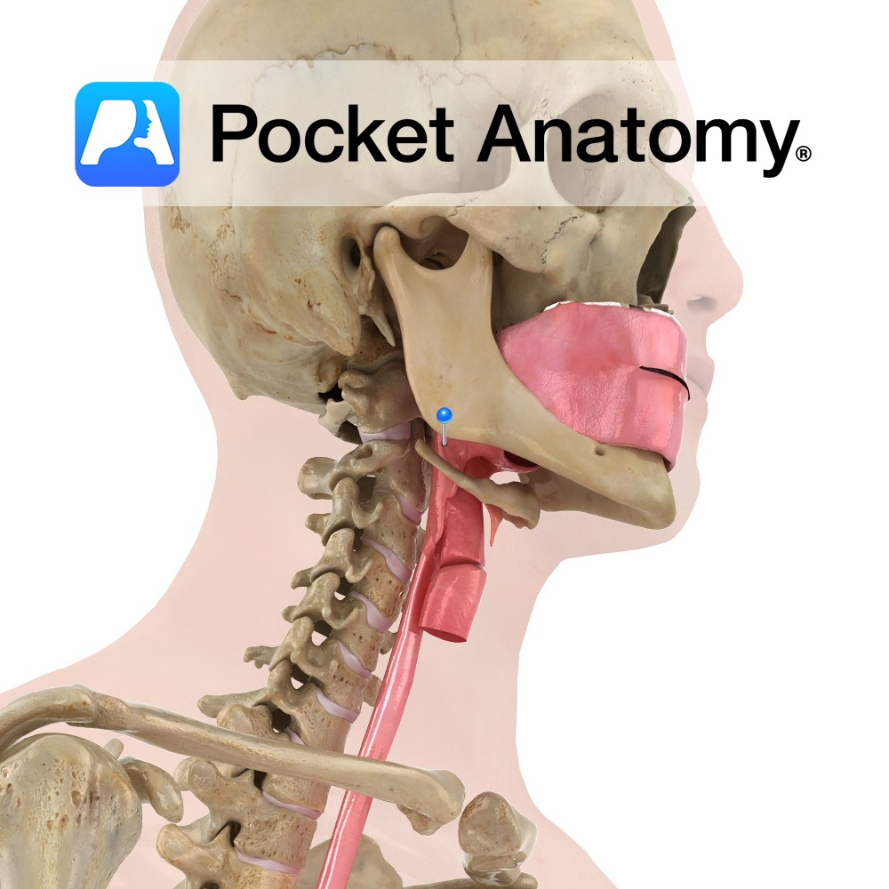
Functions Allows communication between oral and nasal cavity. Separates (with epiglottis) food/drink and air into oesophagus and trachea respectively. Helps (along with nostils and nasal cavity) to warm and humidify inhaled air before lungs. Important area for reverberation when vocalising. Anatomy Cone shaped area at back of throat, typically 3.6cm2 cross section in men, 3.2cm2
- Published in Pocket Anatomy Pins
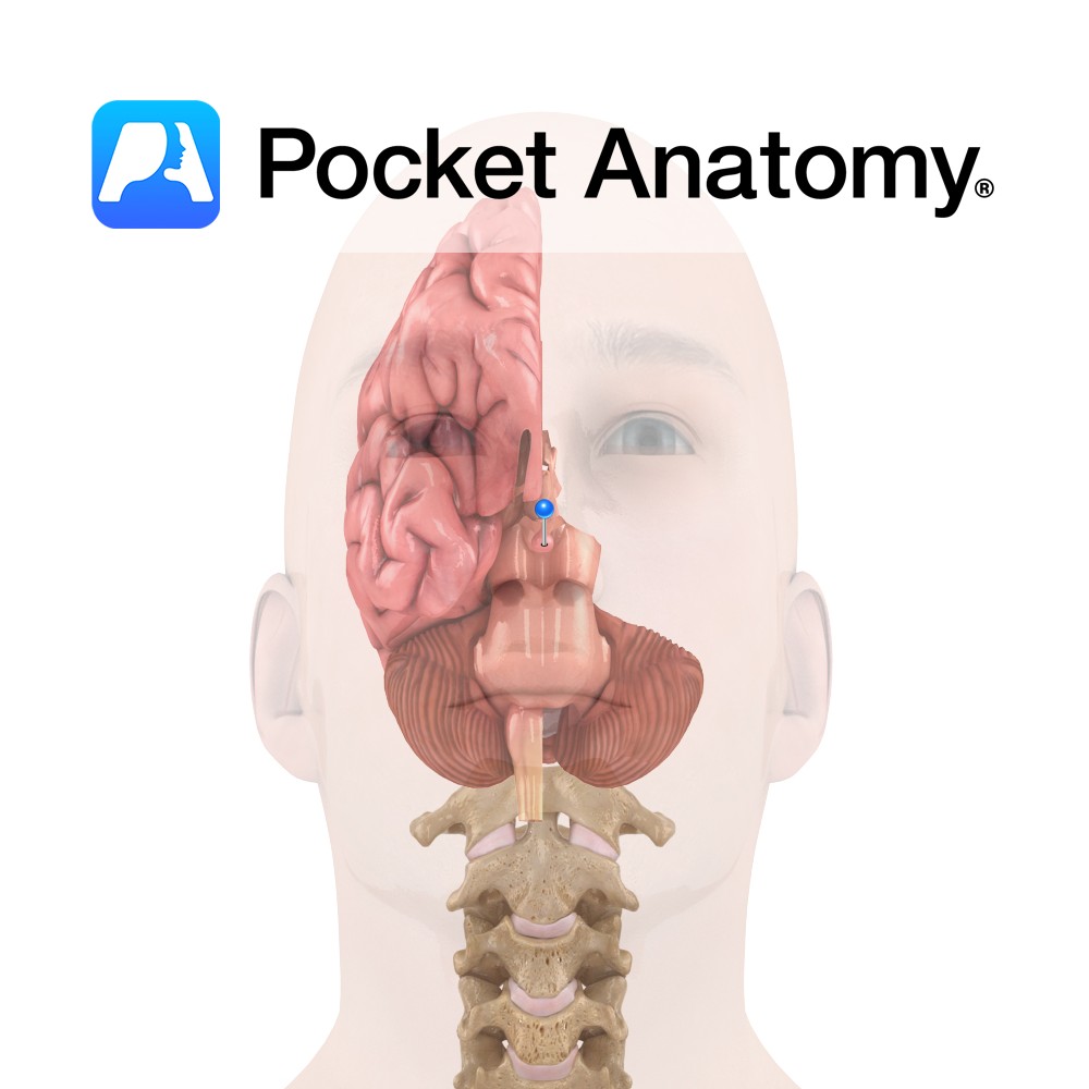
Functions Stores and secretes (anterior lobe also synthesises) a number of essential hormones: Anterior: Human growth hormone, Thyroid-stimulating hormone, Adrenocorticotropic hormone, Beta-endorphin, Prolactin, Luteinising hormone, Follicle-stimulating hormone. Intermediate: Melanocyte-stimulating hormone. Posterior: Antidiuretic hormone (synthesised in supra-optic nucleus), Oxytocin (synthesised in para-ventricular nucleus). Anatomy Endocrine gland located at base of brain; inferior and connected (via infundibular
- Published in Pocket Anatomy Pins