PocketAnatomy® is a registered brand name owned by © eMedia Interactive Ltd, 2009-2022.
iPhone, iPad, iPad Pro and Mac are trademarks of Apple Inc., registered in the U.S. and other countries. App Store is a service mark of Apple Inc.
.jpg)
Anatomy Articulates up with metacarpal at 3rd MCP, down with middle phalanx at proximal interphalangeal joint (PIP). Interested in taking our award-winning Pocket Anatomy app for a test drive?
- Published in Pocket Anatomy Pins
.jpg)
Anatomy Articulates up with metacarpal at 5th MCP, down with middle phalanx at proximal interphalangeal joint (PIP). Clinical Rheumatoid arthritis commoner MCPs and PIPs, osteoarthritis commoner DIPs. Interested in taking our award-winning Pocket Anatomy app for a test drive?
- Published in Pocket Anatomy Pins
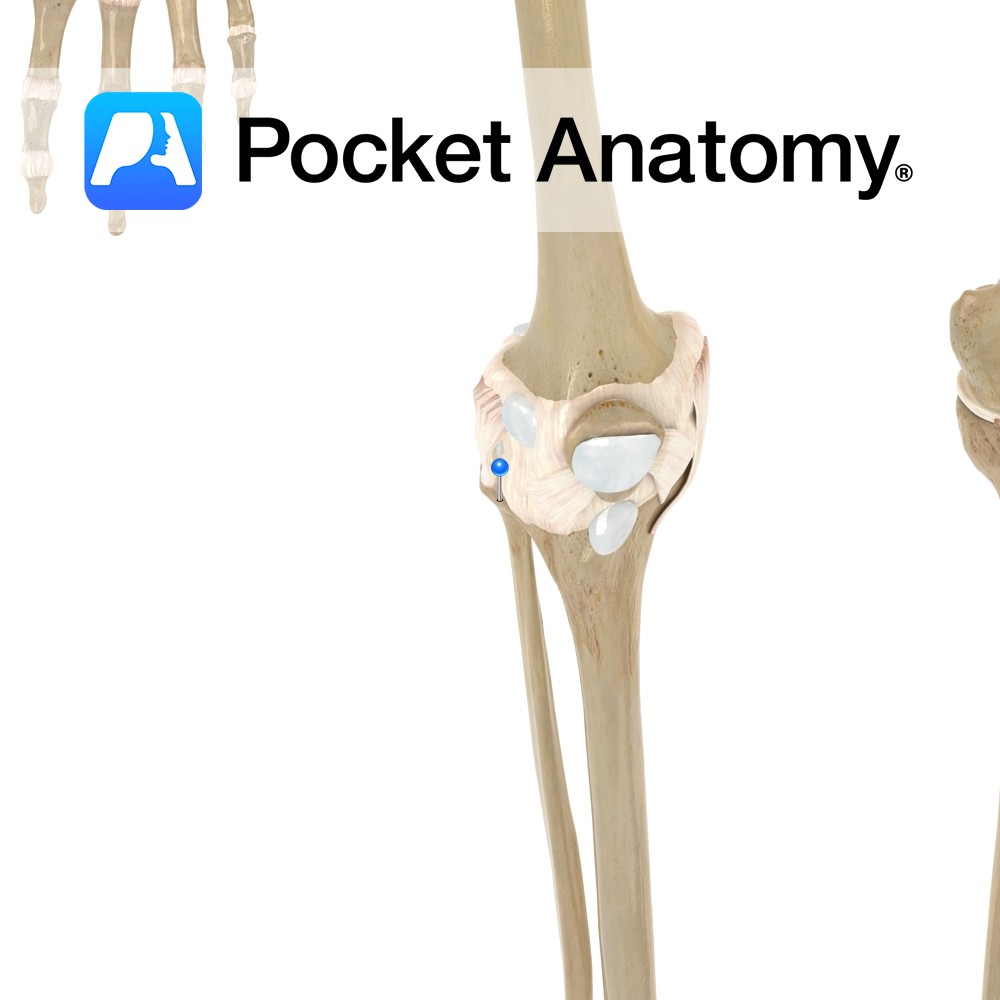
Motion The proximal tibiofibular joint is a synovial plane joint. The lateral condyle of the tibia articulates with the articular facet of the head of the fibula. It allows very little movement. Slight movement occurs to accommodate the movement of the medial malleolus and fibula during plantar or dorsiflexion of the foot. Stability The fibrous
- Published in Pocket Anatomy Pins
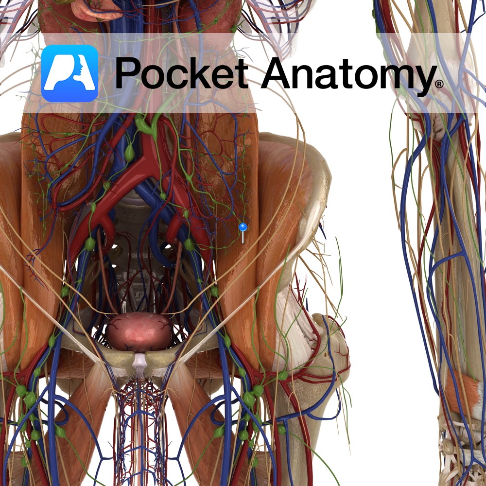
Anatomy Origin: Sides of vertebra T12 to L5 and transverse processes of L1 to L5. Insertion: Lesser trochanter of the femur. Key Relations: -The ureter passes along the medial aspect of the psoas major muscle as it enters the pelvis. -Forms the floor of the femoral triangle. -The femoral nerve (L2 to L4) travels through
- Published in Pocket Anatomy Pins
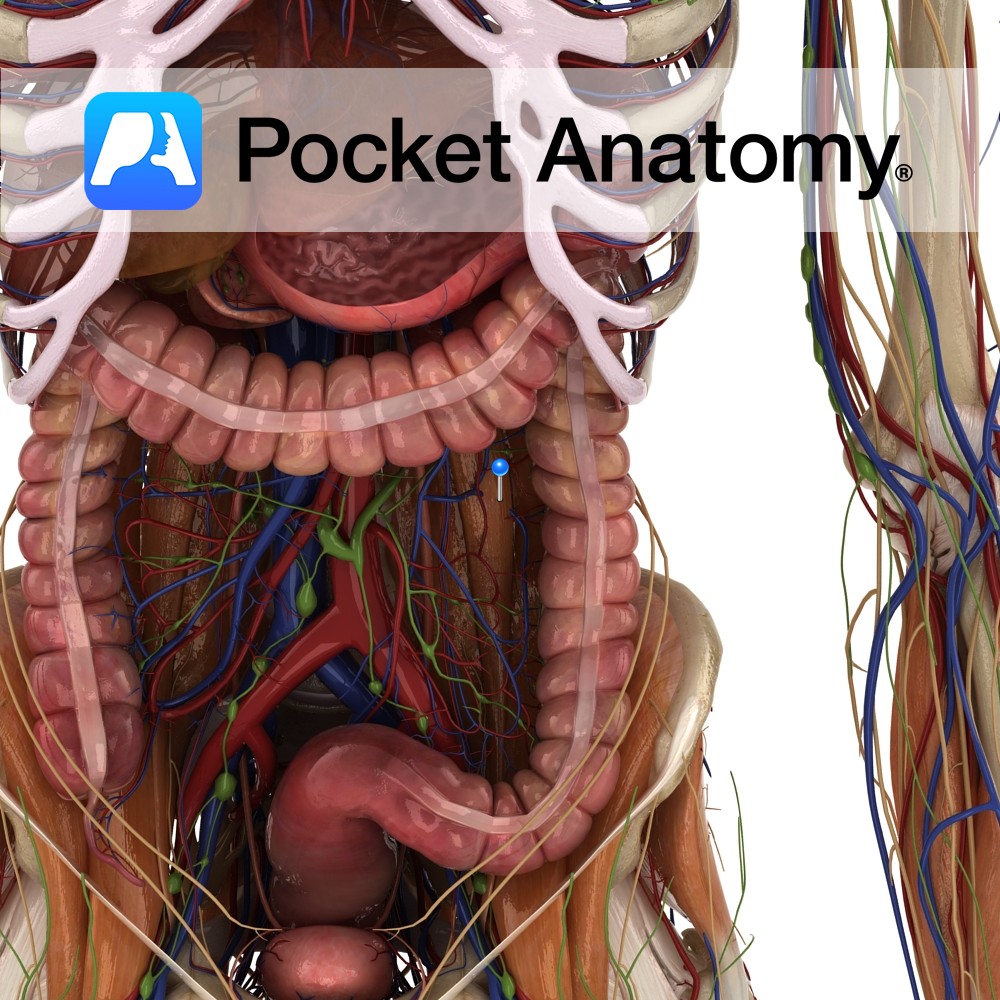
Anatomy Origin: Lateral surface of T12 and L1 and the intervening intervertebral disc. Insertion: Pectineal line of pelvic brim and iliopubic eminence and iliac fascia. Key Relations: -Found in only 60% of population. -When present it is found lying across the surface of the psoas major muscle. Functions Weakly flexes the trunk. Supply Nerve Supply:
- Published in Pocket Anatomy Pins

Anatomy Forms part of the border of obturator foramen, joins with inferior ramus of ischium. Vignette Ramus (Latin); branch. Interested in taking our award-winning Pocket Anatomy app for a test drive?
- Published in Pocket Anatomy Pins
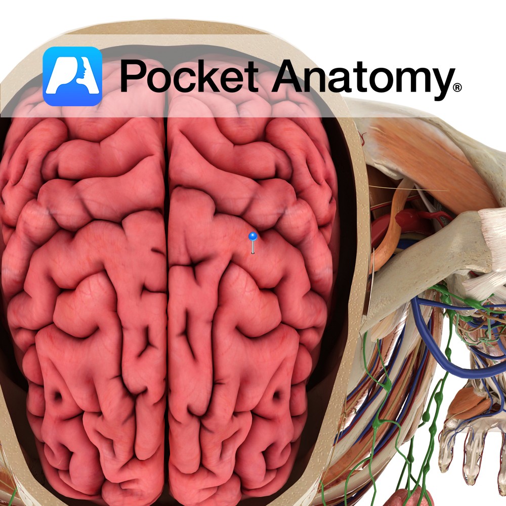
Anatomy A gyrus located anterior to the central sulcus. It extends anteroinferiorly from the great longitudinal fissure and ends just above the lateral sulcus. It forms part of the frontal lobe. Blood supply: Supplied by the middle cerebral artery, and medially by the anterior cerebral artery, to a lesser extent. Functions Is the location of
- Published in Pocket Anatomy Pins
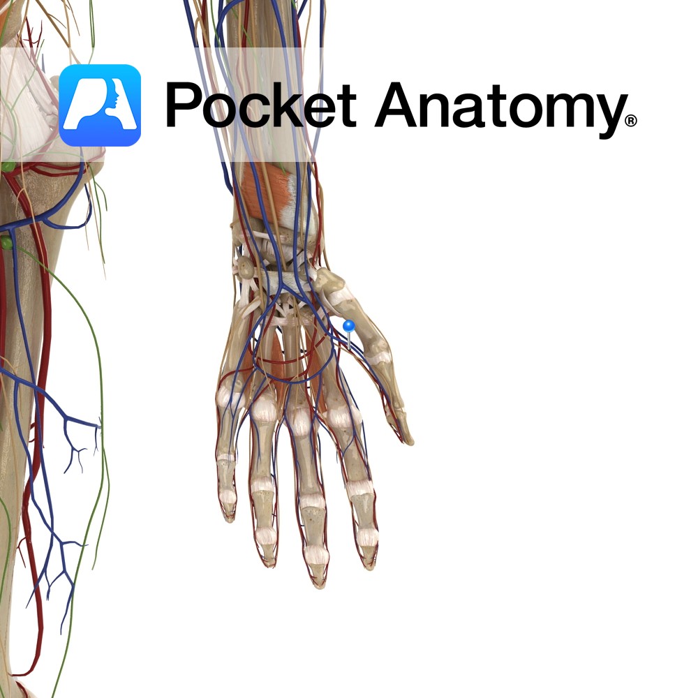
Anatomy Course Branches from the radial artery, just as the radial artery is veering medially to become the deep palmar arterial arch. It travels towards the thumb between the first dorsal interosseous artery and the adductor pollicis muscle. Supply Main blood supply to the thumb. Interested in taking our award-winning Pocket Anatomy app for a
- Published in Pocket Anatomy Pins
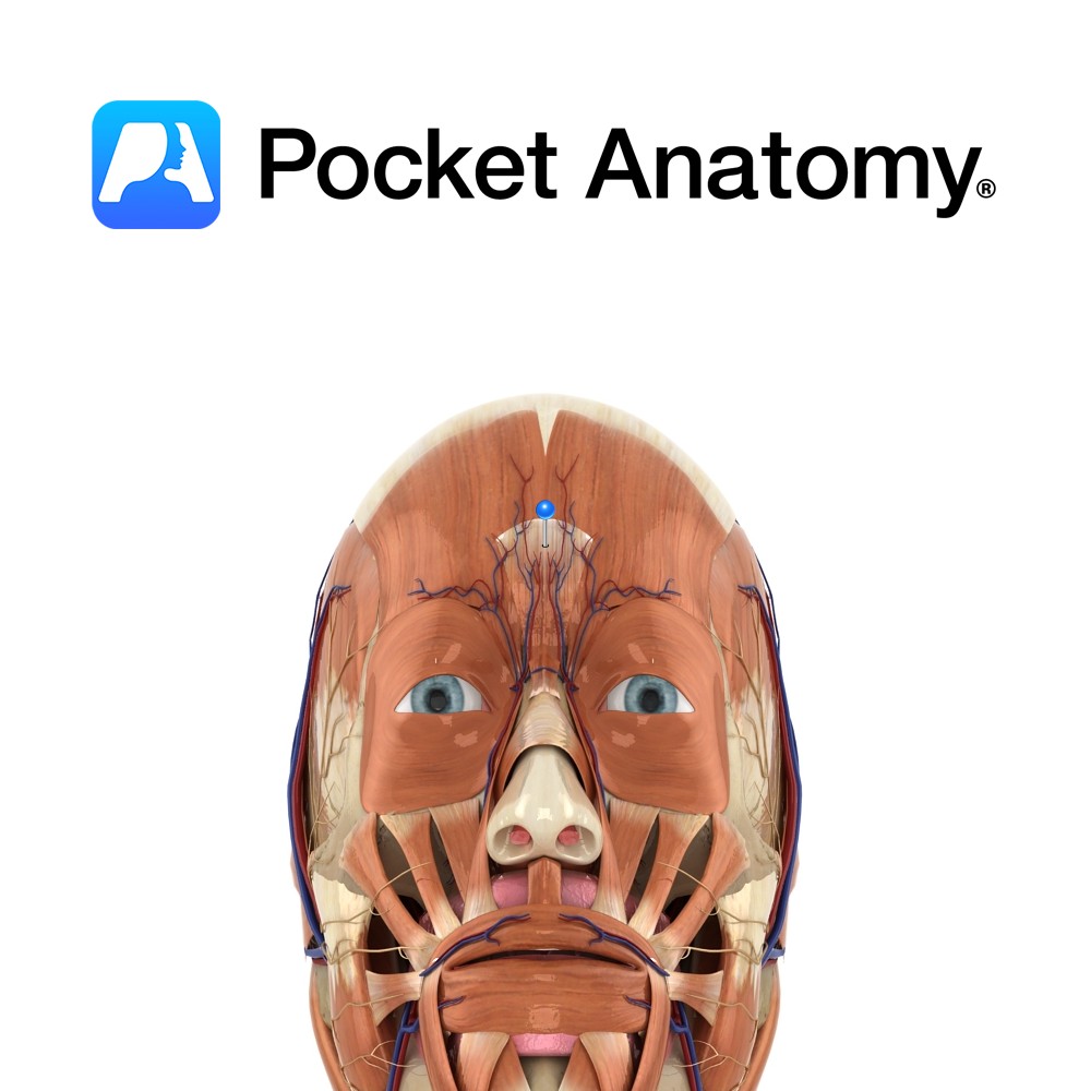
Anatomy Origin: Nasal bone and upper part of the lateral nasal cartilage. Insertion: Skin of lower forehead between the eyebrows. Key Relations: Lies over the nasal bone. Functions Draws medial border of eyebrows inferiorly resulting in transverse wrinkles over the bridge of the nose. e.g. as in an expression of anger.. Supply Nerve Supply: Zygomatic
- Published in Pocket Anatomy Pins
-artery.jpg)
Anatomy Course Branches from the brachial artery about a third of the way along the humerus. It enters the posterior compartment of the arm with the radial nerve, passing through the space made by the lateral margin of long head of the triceps, the inferior margin of teres major and the shaft of the humerus.
- Published in Pocket Anatomy Pins