PocketAnatomy® is a registered brand name owned by © eMedia Interactive Ltd, 2009-2022.
iPhone, iPad, iPad Pro and Mac are trademarks of Apple Inc., registered in the U.S. and other countries. App Store is a service mark of Apple Inc.
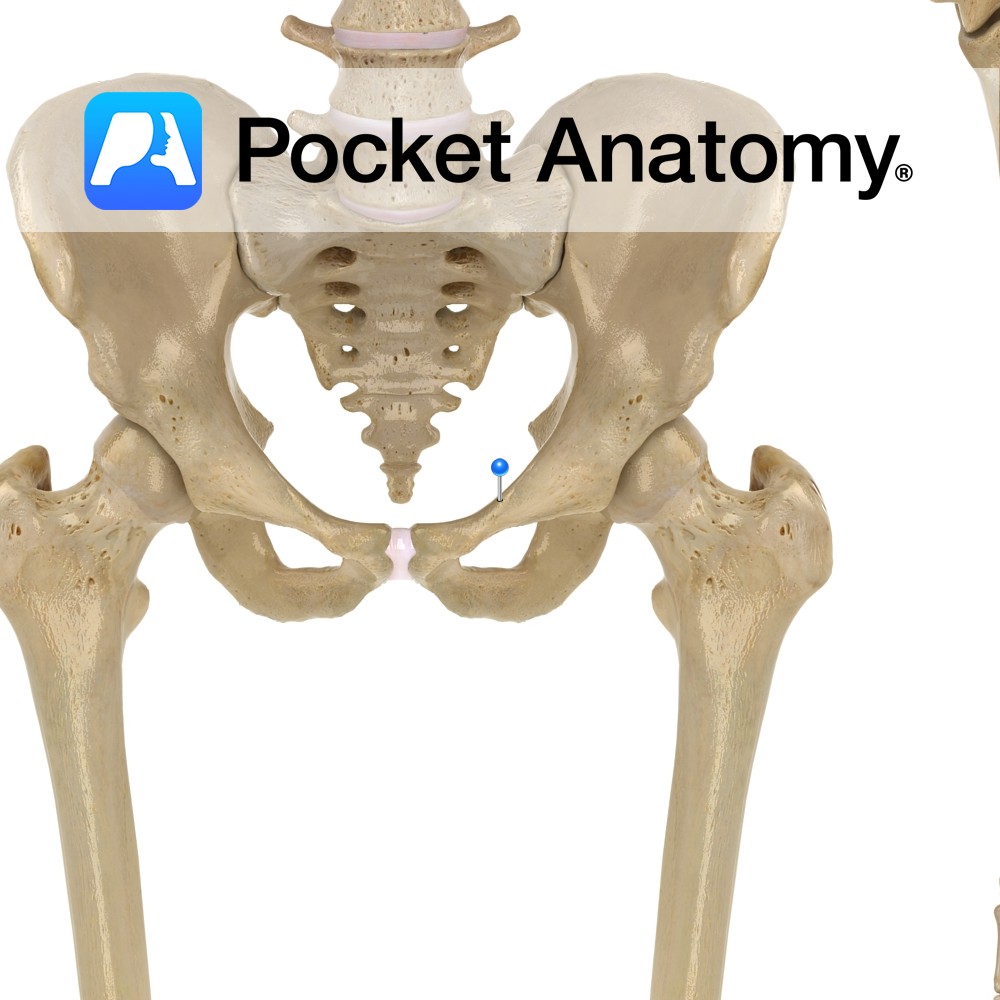
Anatomy Also known as pectin pubis. Ridge on upper inner aspect superior ramus, continuous with arcuate line of ilium (together called the iliopectineal line); part of pelvic brim. Interested in taking our award-winning Pocket Anatomy app for a test drive?
- Published in Pocket Anatomy Pins
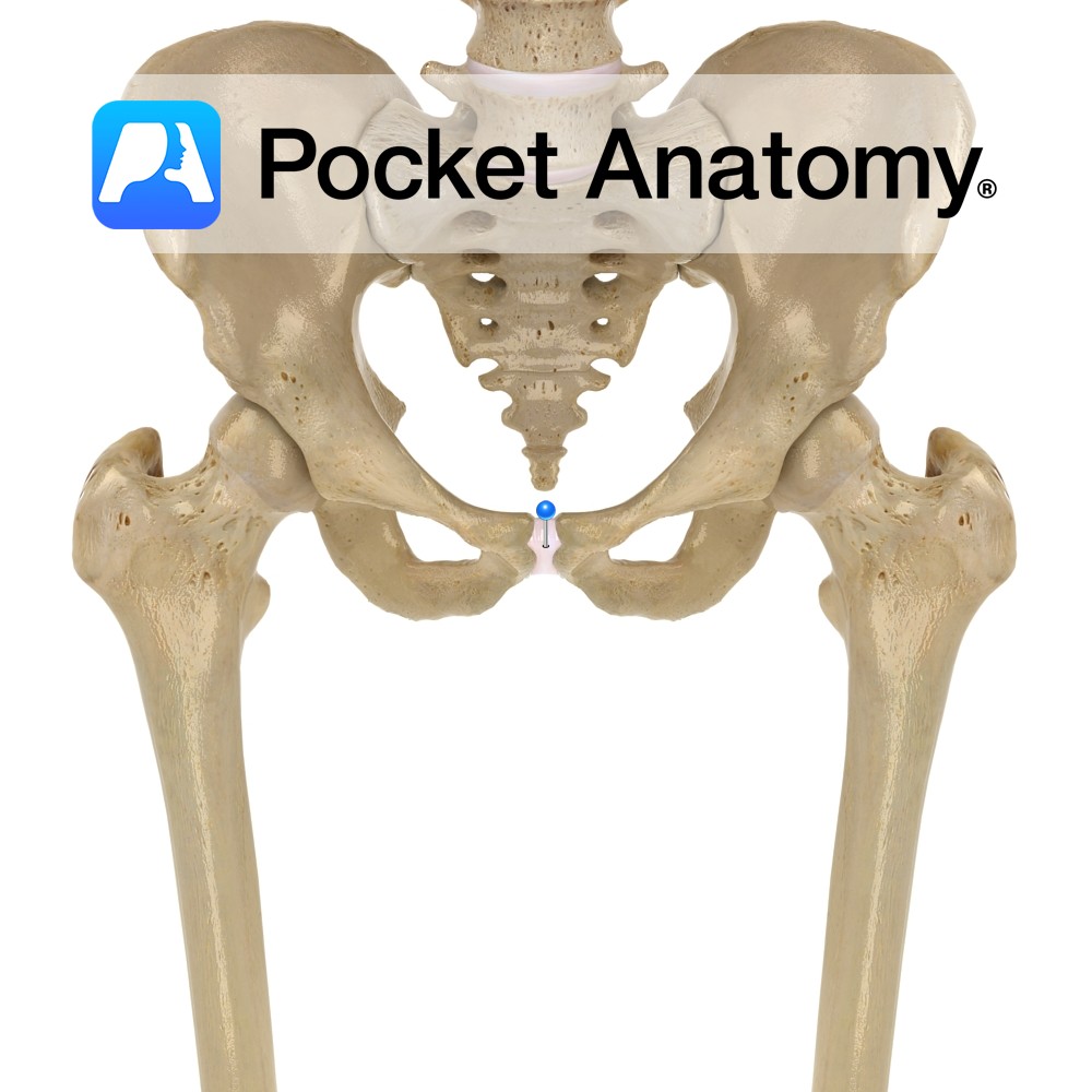
Anatomy Midline cartilaginous joint between superior pubic rami (left and right). Clinical In pregnancy, there is hormone-mediated laxity of the symphysis, assisting passage of baby during birth; the joint may disrupt (diastasis) leading to pelvic girdle pain. Ankylosing spondylitis causes bony union. Interested in taking our award-winning Pocket Anatomy app for a test drive?
- Published in Pocket Anatomy Pins
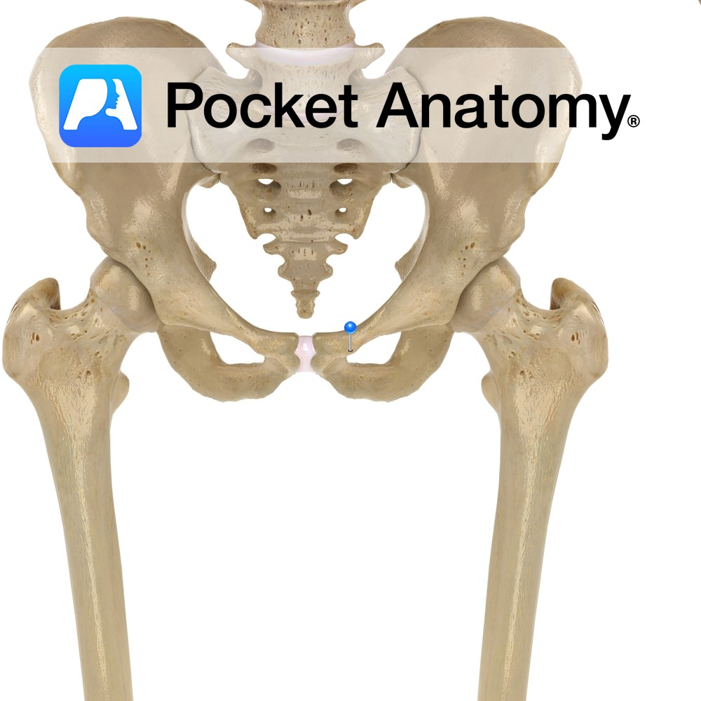
Anatomy Small prominent forward-projecting bulge on medial part of upper surface of superior ramus of pubis, to which the inguinal ligament is attached. Clinical Pectineal line starts at tubercle. Interested in taking our award-winning Pocket Anatomy app for a test drive?
- Published in Pocket Anatomy Pins
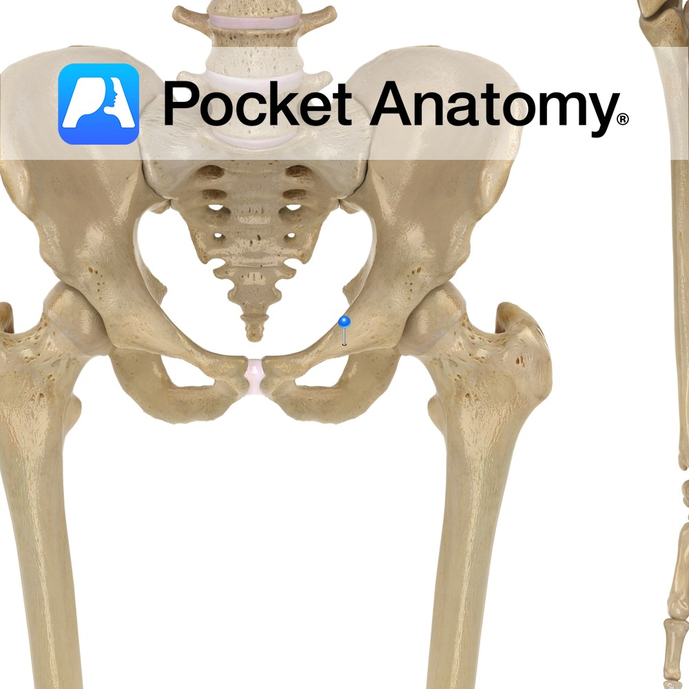
Anatomy Forms part of border of obturator foramen. Left and right pubic rami join at pubic symphysis. Clinical Easily palpated. Just in front of bladder and above external genitalia. Interested in taking our award-winning Pocket Anatomy app for a test drive?
- Published in Pocket Anatomy Pins
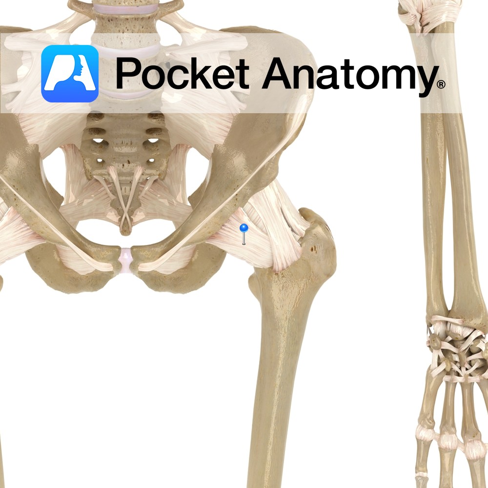
Anatomy Attaches from just medial to the iliopubic eminence, adjacent bone and obturator membrane. It blends with the joint capsule and the iliofemoral ligament to attach to the femur. Functions Provides static support to the hip joint. Interested in taking our award-winning Pocket Anatomy app for a test drive?
- Published in Pocket Anatomy Pins
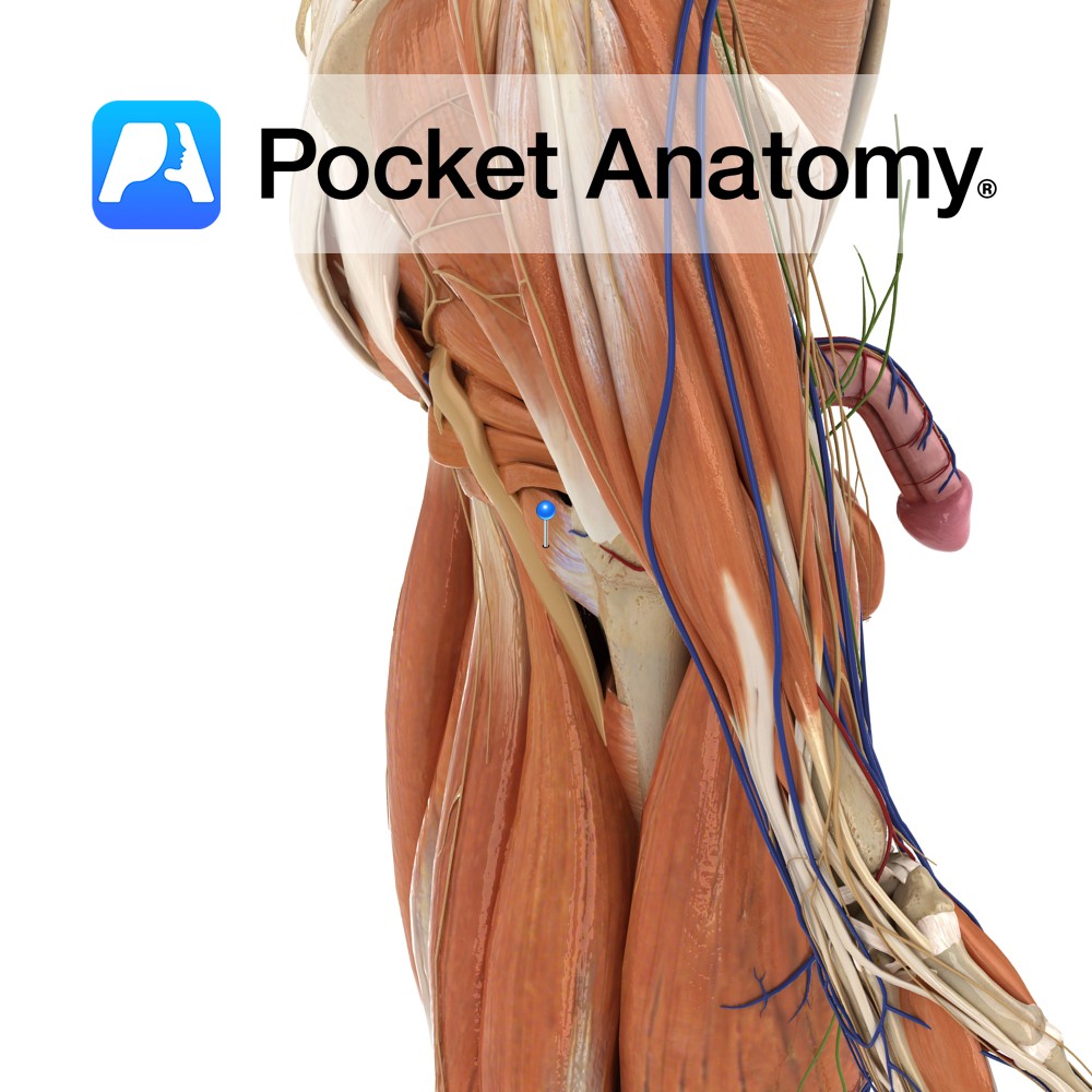
Anatomy Origin: Superior lateral margin of the ischial tuberosity. Insertion: Quadrate tubercle on the intertrochanteric crest of the femur. Key Relations: Lies between the inferior gemellus and the upper margin of the adductor magnus muscle. Functions Laterally rotates and adducts the thigh at the hip joint. Supply Nerve Supply: Nerve to quadratus femoris (L5, S1).
- Published in Pocket Anatomy Pins
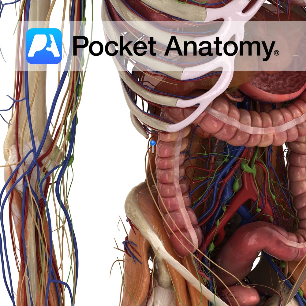
Anatomy Origin: Transverse processes of L3 to L5, iliolumbar ligament and iliac crest. Insertion: Inferior border of 12th rib and transverse processes of L1 to L3. Key relations: -Quadratus lumborum lies deep to the colon, kidney, psoas major, psoas minor and the diaphragm. -The subcostal, iliohypogastric and ilioinguinal nerveslie anterior to the fascia that overlies
- Published in Pocket Anatomy Pins
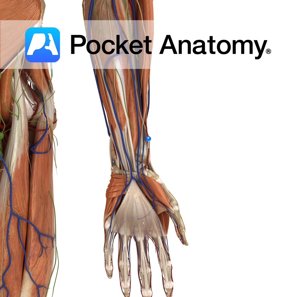
Anatomy Course Originates from the brachial artery at the head of the radius, in the cubital fossa at the elbow. It runs along the lateral aspect of the forearm, passing deep to the brachioradialis muscle, running with a superficial branch of the radial nerve. It passes lateral to the flexor carpi radialis tendon and winds
- Published in Pocket Anatomy Pins
.jpg)
Anatomy Articulates posteriorly with 5th metatarsal, anteriorly with 5th middle phalanx. Interested in taking our award-winning Pocket Anatomy app for a test drive?
- Published in Pocket Anatomy Pins
.jpg)
Anatomy At near end is 1st MCP. Thumb has only 2 phalanges, joint between is intermediate interphalangeal. Interested in taking our award-winning Pocket Anatomy app for a test drive?
- Published in Pocket Anatomy Pins