PocketAnatomy® is a registered brand name owned by © eMedia Interactive Ltd, 2009-2022.
iPhone, iPad, iPad Pro and Mac are trademarks of Apple Inc., registered in the U.S. and other countries. App Store is a service mark of Apple Inc.
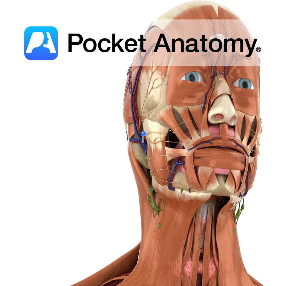
Anatomy Course Formed when the superficial temporal vein and the maxillary vein unite, just superior to the parotid gland. It descends through the parotid gland before dividing into an anterior and posterior branch. Drain The retromandibular vein contributes to the drainage of the deep facial structures and the temporal region of the scalp. Interested in
- Published in Pocket Anatomy Pins
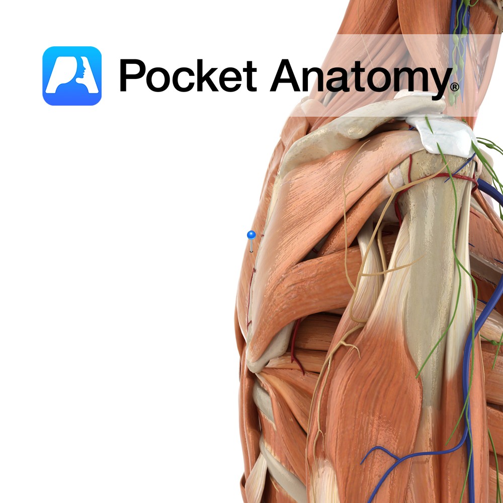
Anatomy Origin: Spinous processes of T2 to T5. Insertion: Medial border of the scapula between the spine and the inferior angle, inferior to the attachment of rhomboid minor. Key Relations: -Inferior to rhomboid minor. -Posterior to trapezius except at the triangle of auscultation. Functions -Retracts the scapula e.g. pulling open a drawer. -Working with the
- Published in Pocket Anatomy Pins
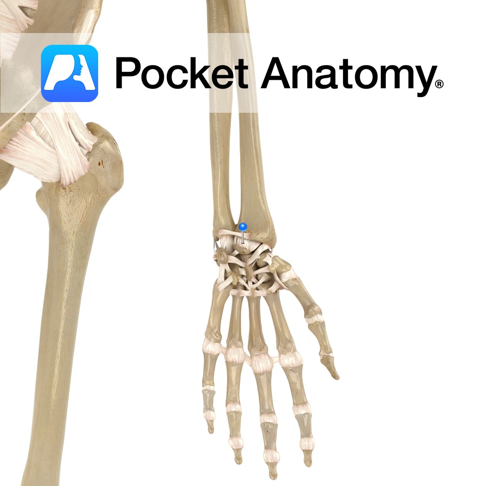
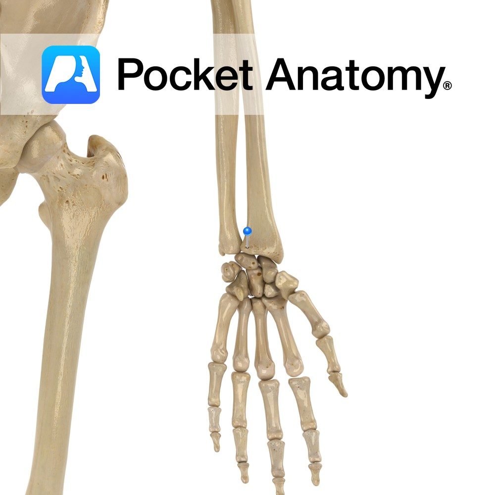
Anatomy Attaches from palmar surface of the radius between the lunate and scaphoid fossae. It blends with the scapholunate interosseous ligament. Functions It provides palmar stability to the wrist and carpal joints. Interested in taking our award-winning Pocket Anatomy app for a test drive?
- Published in Pocket Anatomy Pins
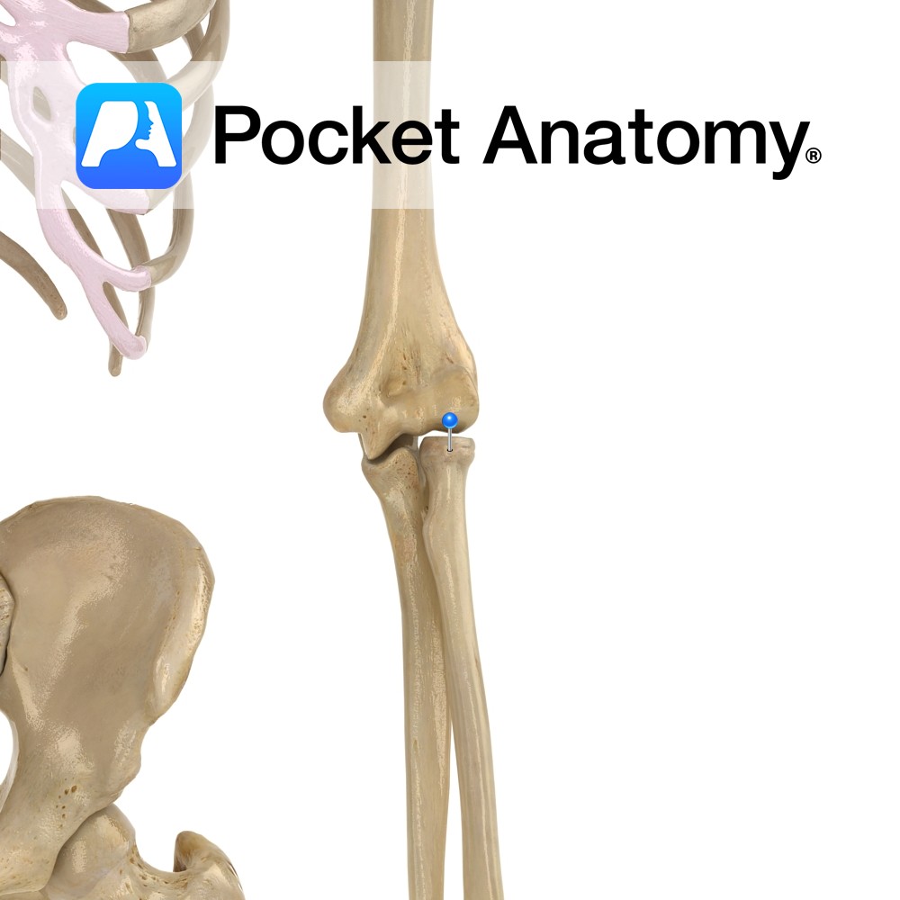
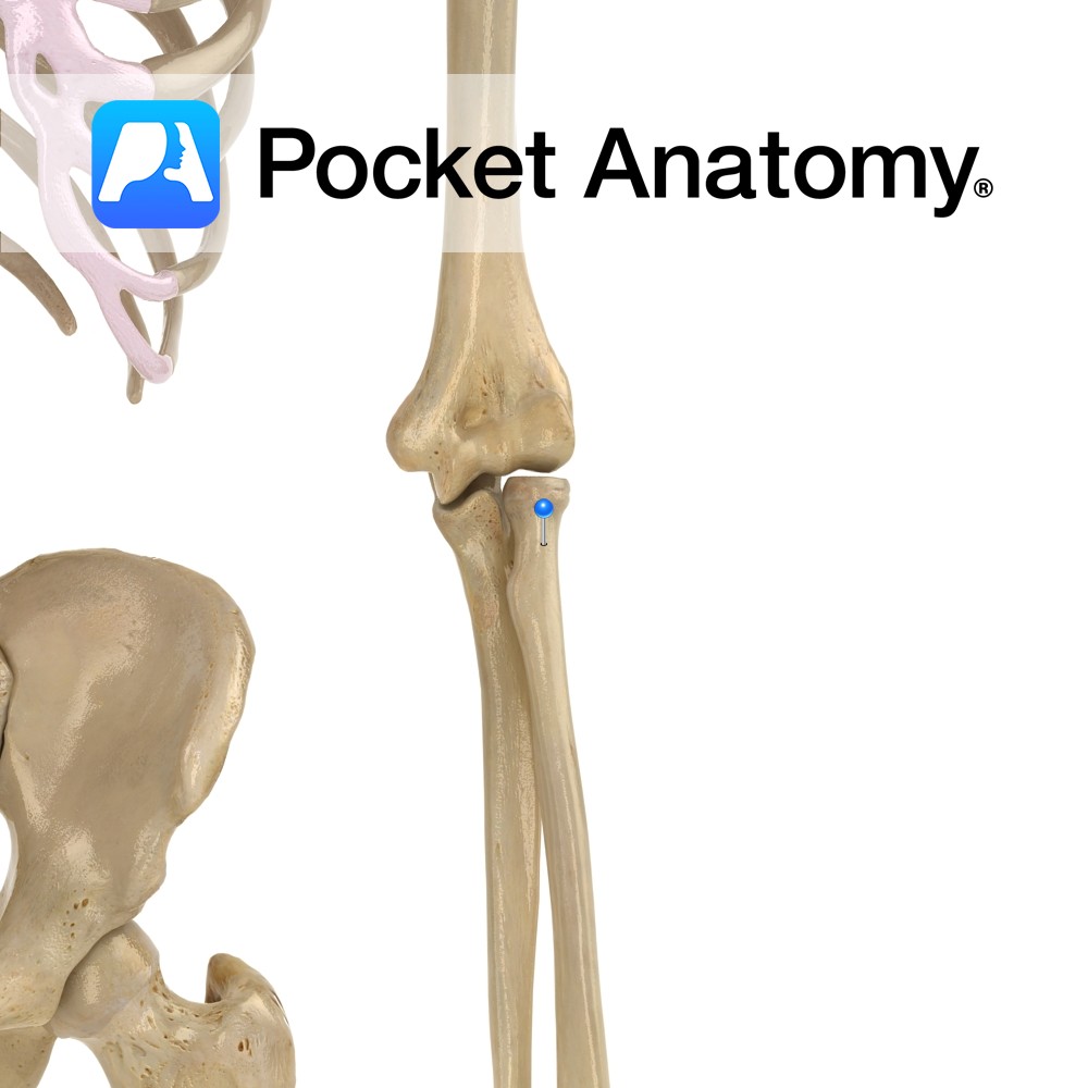
Anatomy Almost cylindrical, articulates with humerus at capitulum, and ulna at radial notch (proximal radioulnar joint). Radiohumeral joint is hinge; movement is flexion-extension. Proximal radioulnar joint is pivot; movement is rotation of radial head inside ring formed by radial notch of ulna and annular ligament. Clinical Radial head is proximal and articulates with a notch
- Published in Pocket Anatomy Pins
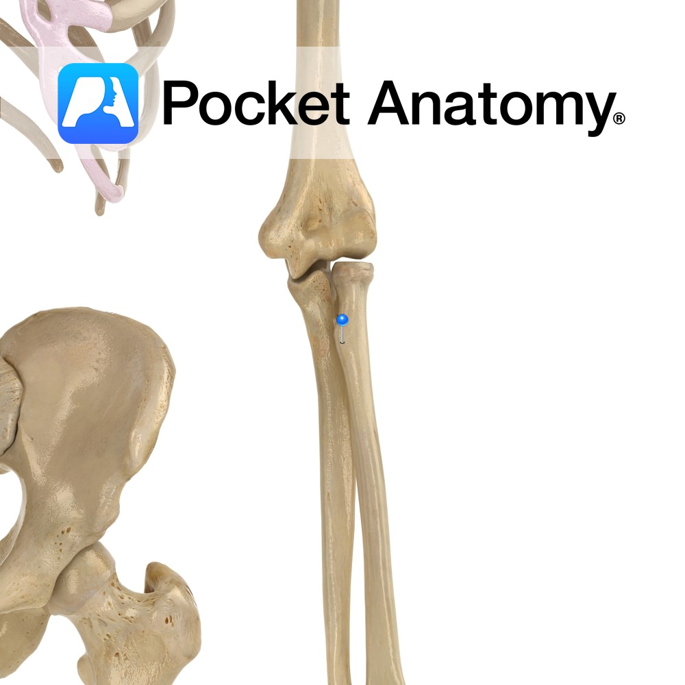
Anatomy A narrowing below the radial head; part of supinator inserts behind. Interested in taking our award-winning Pocket Anatomy app for a test drive?
- Published in Pocket Anatomy Pins
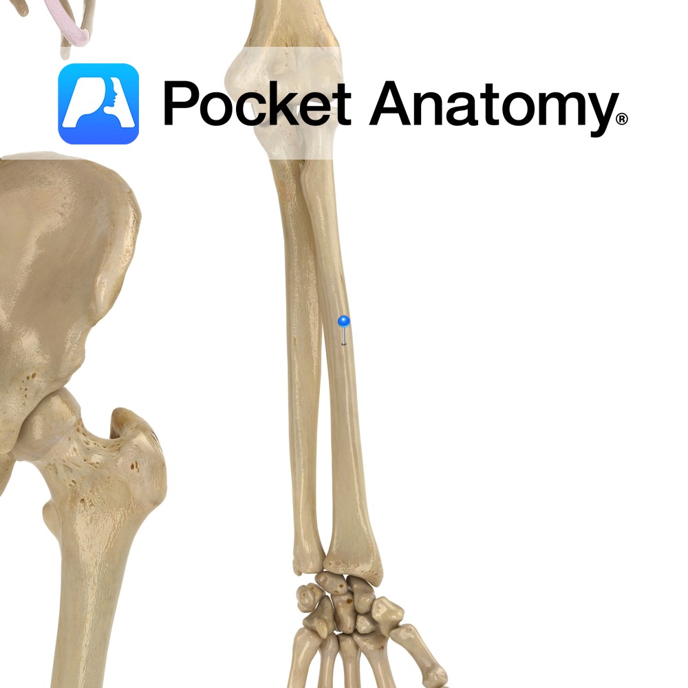
Anatomy A bulge medially, just below radial neck, for insertion of tendon biceps brachii. Interested in taking our award-winning Pocket Anatomy app for a test drive?
- Published in Pocket Anatomy Pins
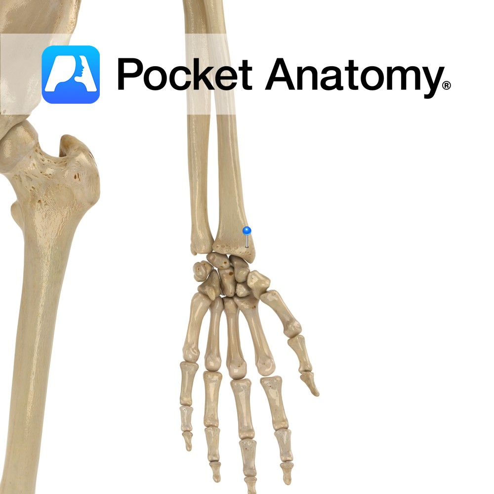
Anatomy Broader towards the bottom, slightly curved convex laterally. Radius and ulna joined (syndesmosis joint – allows slight movement) by interosseous membrane along their lengths. Clinical Dinnerfork or bayonet deformity in Colles’ (commonest wrist) fracture; a fall on the outstretched hand). Radial pulse felt against thumb-side anterior/palmar aspect, lower quarter. Interested in taking our award-winning
- Published in Pocket Anatomy Pins
Anatomy Bulge down from lateral side (thumb-side) lower extremity, conical in shape, giving attachment to tendons of brachoradialis, abductor pollicis longus and extensor pollicis brevis, and radial collateral ligament of wrist. Bigger, more inferior/distal than ulnar styloid. Vignette Chauffeur’s fracture; pressure of scaphoid on styloid. Interested in taking our award-winning Pocket Anatomy app for a
- Published in Pocket Anatomy Pins
Anatomy Almost cylindrical head of ulna articulates with ulnar notch of radius, distal radioulnar joint. A pivot joint; movement is rotation of radius (and hand; ulna main contributor to elbow joint, radius to wrist joint) around the ulnar head. Clinical Radial head is proximal and articulates with a notch on the ulna (proximal radioulnar joint
- Published in Pocket Anatomy Pins
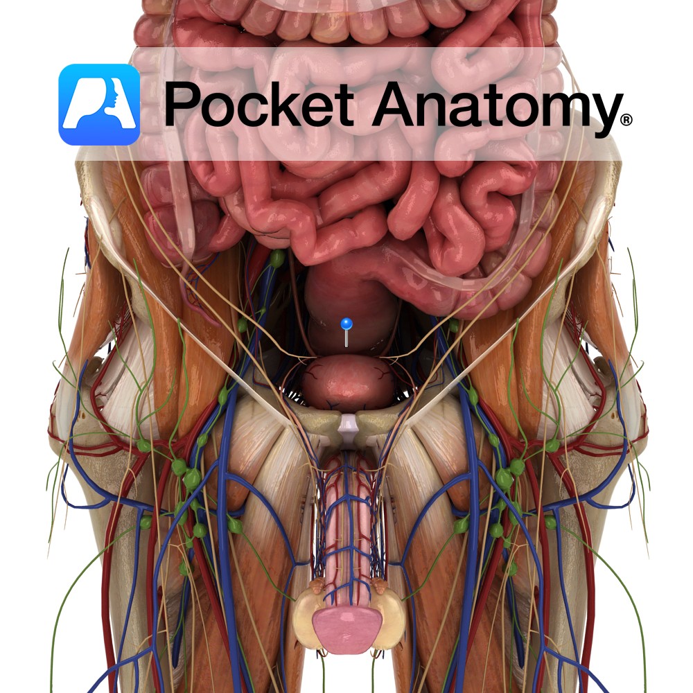
Anatomy Continuous with sigmoid colon above and anal canal below, sitting in and following sacrococcygeal curve and on to c 2.5 cms below coccyx, to level of apex prostate, then turning back abruptly and dilating (rectal ampulla) to anal canal. 3 or 4 transverse folds (Houston’s valves) overlap when rectum empty and both support weight
- Published in Pocket Anatomy Pins