PocketAnatomy® is a registered brand name owned by © eMedia Interactive Ltd, 2009-2022.
iPhone, iPad, iPad Pro and Mac are trademarks of Apple Inc., registered in the U.S. and other countries. App Store is a service mark of Apple Inc.
-%5Btrue-rib%5D.jpg)
Anatomy Only rib with sternal articulation that straddles manubrium and body. Interested in taking our award-winning Pocket Anatomy app for a test drive?
- Published in Pocket Anatomy Pins
-%5Btrue-rib%5D.jpg)
Anatomy From 6th rib down, the costal cartilages (CC) above receive contribution from CC below. Interested in taking our award-winning Pocket Anatomy app for a test drive?
- Published in Pocket Anatomy Pins
-%5Bfalse-rib%5D.jpg)
Anatomy False rib, i.e. articulates posteriorly but not directly anteriorly. Its costal cartilages (CC) merges with CC of 7th rib above. Interested in taking our award-winning Pocket Anatomy app for a test drive?
- Published in Pocket Anatomy Pins
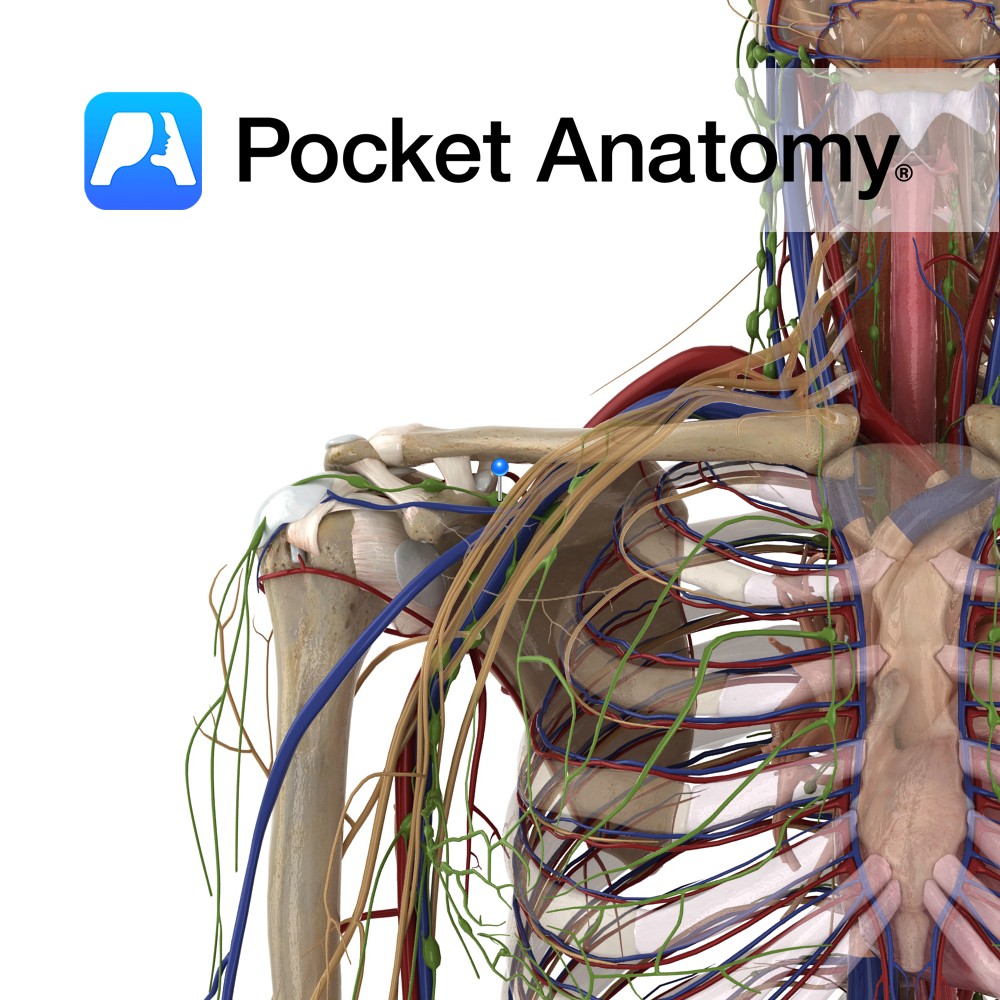
Anatomy Drains R Upper Limb, R side of head and neck and R side of Thorax/chest (and sometimes L lung lower lobe), formed by union of 3 Trunks (R Jugular, R Bronchomediastinal and R Subclavian), about 1/2 inch in length, empties into R Subclavian Vein. Clinical 85% of what leaves through the arteriolar end of
- Published in Pocket Anatomy Pins
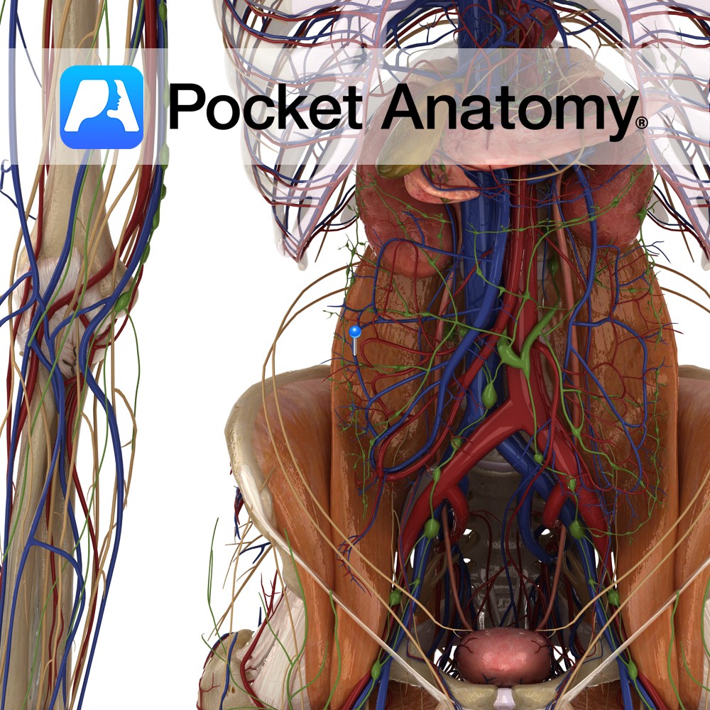
Anatomy Course Begins from a series of veins that drain the ascending colon. It travels through the mesentery to drain into the superior mesenteric vein. Drain Drains the ascending colon. Interested in taking our award-winning Pocket Anatomy app for a test drive?
- Published in Pocket Anatomy Pins
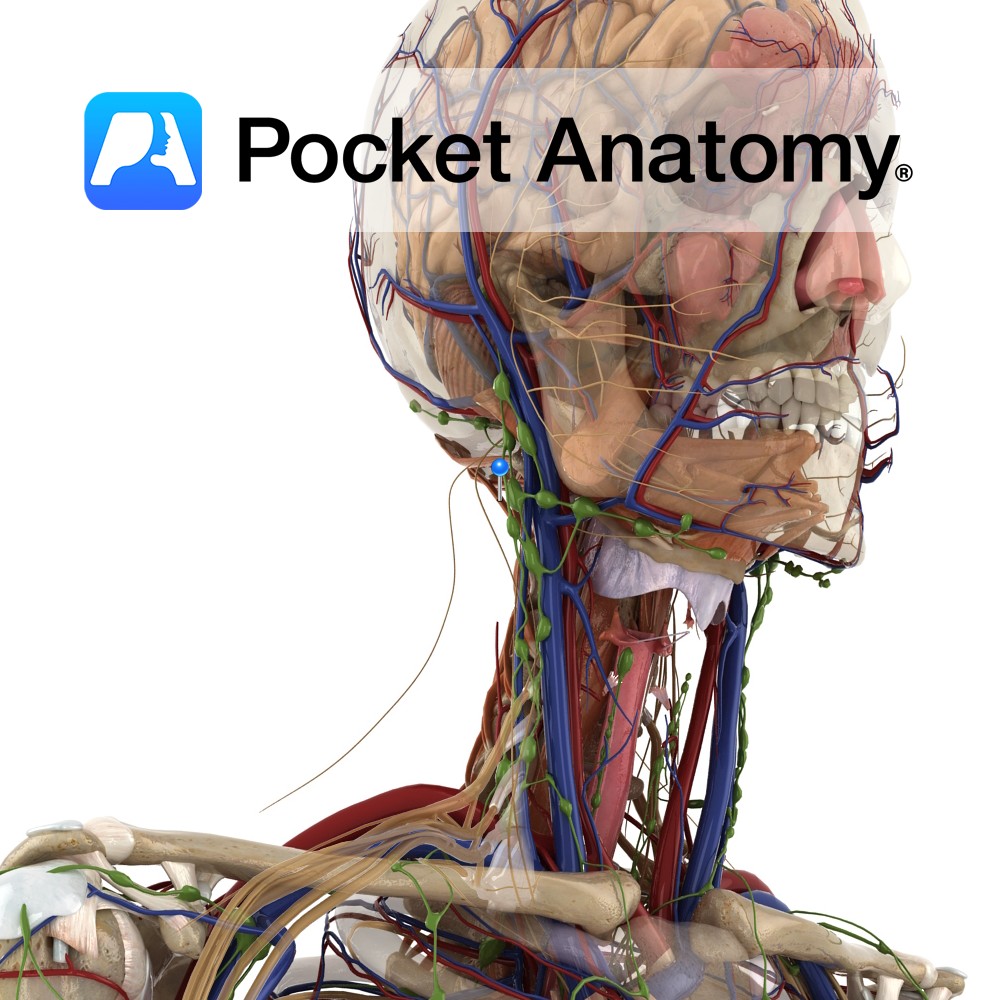
Anatomy Origin: Spinous process of axis. Insertion: Middle third of inferior nuchal line. Key Relations: Forms the superior and medial border of the suboccipital triangle. Functions -Acts bilaterally to extend the head at the atlanto-occipital joint. -Acts unilaterally to rotate the head to the same side at the atlanto-occipital joint. Supply Innervation: Suboccipital nerve (C1).
- Published in Pocket Anatomy Pins
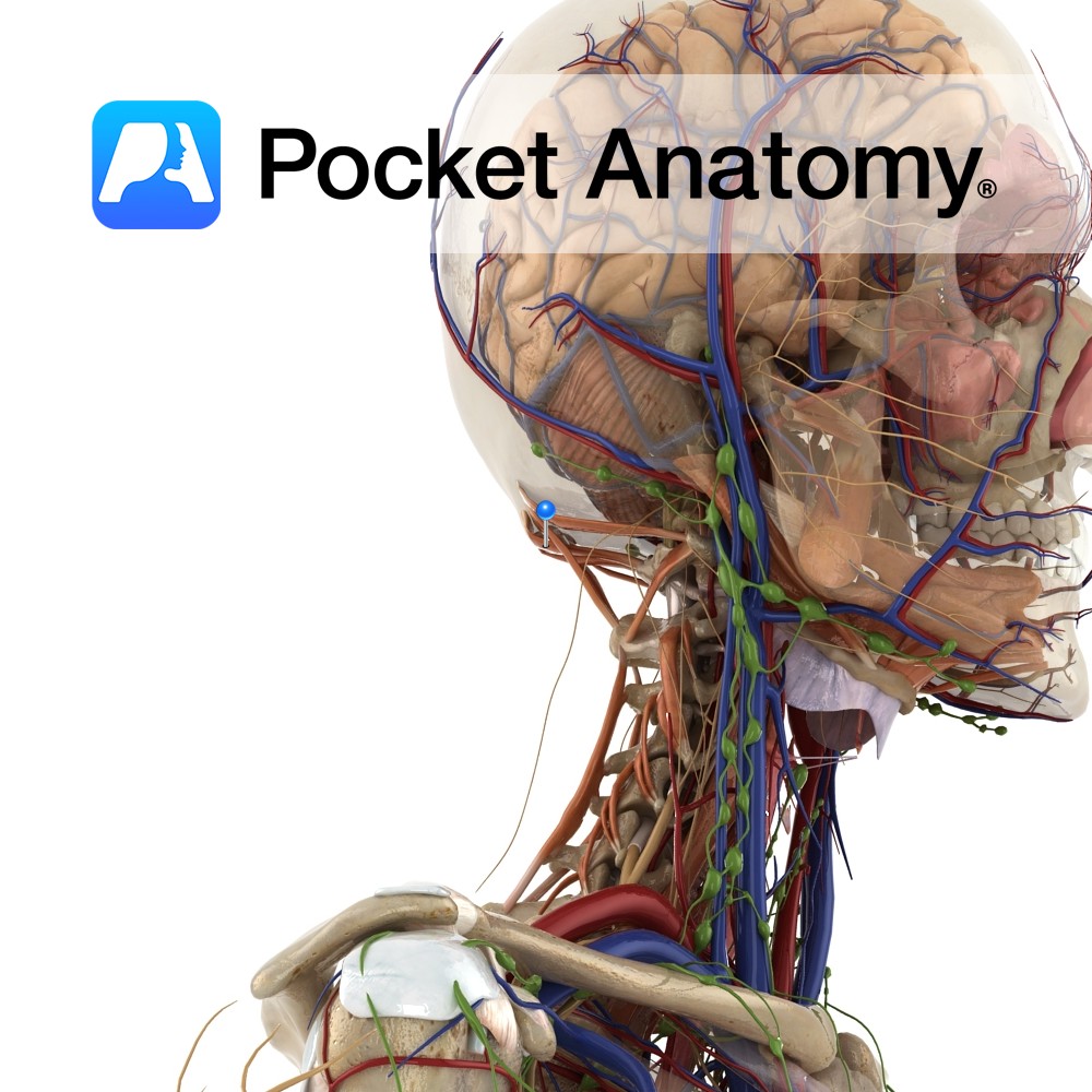
Anatomy Origin: posterior tubercle of atlas. Insertion: From foramen magnum to medial third of inferior nuchal line. Functions Acts bilaterally to extend the head at the atlanto-occipital joint. Supply Innervation: Suboccipital nerve (C1). Blood supply: Muscular branches of vertebral artery. Clinical Slips of the muscle attach to the spinal dura and may be involved in
- Published in Pocket Anatomy Pins
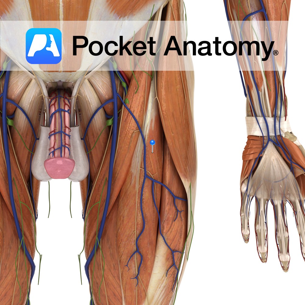
Anatomy Origin: Straight head: anterior inferior iliac spine. Reflected head: a groove on the upper surface of the acetabulum and from the fibrous capsule of the hip joint. Insertion: Base of the patella via the quadriceps femoris tendon. The quadriceps femoris tendon is functionally continuous with the patellar ligament which runs from the apex of
- Published in Pocket Anatomy Pins
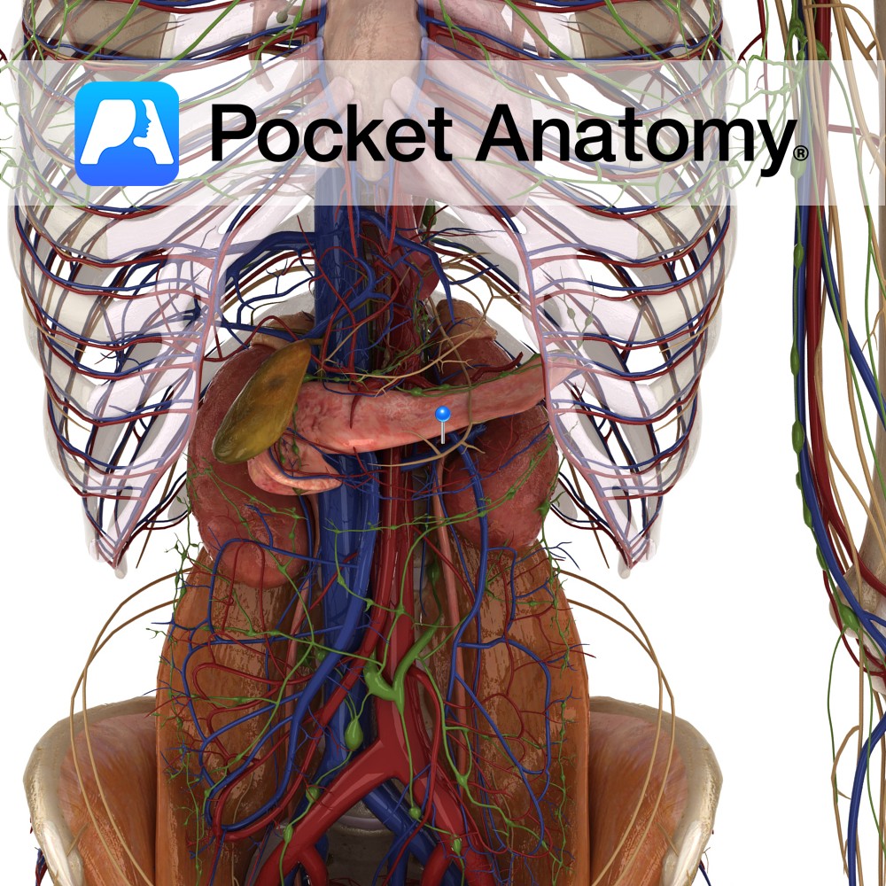
Anatomy Course The renal arteries branch directly from the abdominal aorta at approximately vertebral level L1-L2. The right renal artery is longer and must pass behind the inferior vena cava to reach the right kidney. The left renal artery usually originates slightly higher. Both travel horizontally and retroperitoneally to their respective kidneys. Supply Supply blood
- Published in Pocket Anatomy Pins
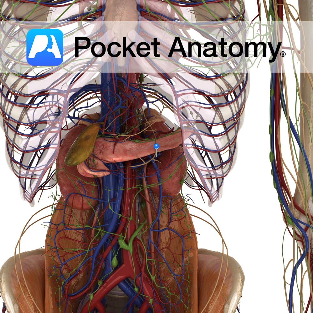
Anatomy Course Begin at the hilums of the kidneys, running horizontally and retroperitoneally to the inferior vena cava. They drain into the inferior vena cava at the approximate vertebral level of L2. Because the left renal vein is longer, and must cross the aorta, it receives a number of tributaries, such as the left phrenic
- Published in Pocket Anatomy Pins