PocketAnatomy® is a registered brand name owned by © eMedia Interactive Ltd, 2009-2022.
iPhone, iPad, iPad Pro and Mac are trademarks of Apple Inc., registered in the U.S. and other countries. App Store is a service mark of Apple Inc.
Anatomy Lowest part of spinal (vertebral) canal, in which spinal cord courses, down to level L1-2, below which, sacral nerves descend into sacral canal as part of cauda equinae. Vignette Cauda Equinae (Latin); horse’s tail. Interested in taking our award-winning Pocket Anatomy app for a test drive?
- Published in Pocket Anatomy Pins

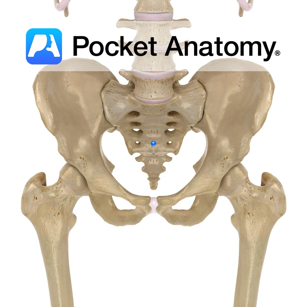
Anatomy Whereas in the other vertebrae the laminae meet and join (and from their juncture a spinous process arises), the S5 (and sometimes S4) laminae do not, leaving a gap or hiatus or natural fissure. Interested in taking our award-winning Pocket Anatomy app for a test drive?
- Published in Pocket Anatomy Pins
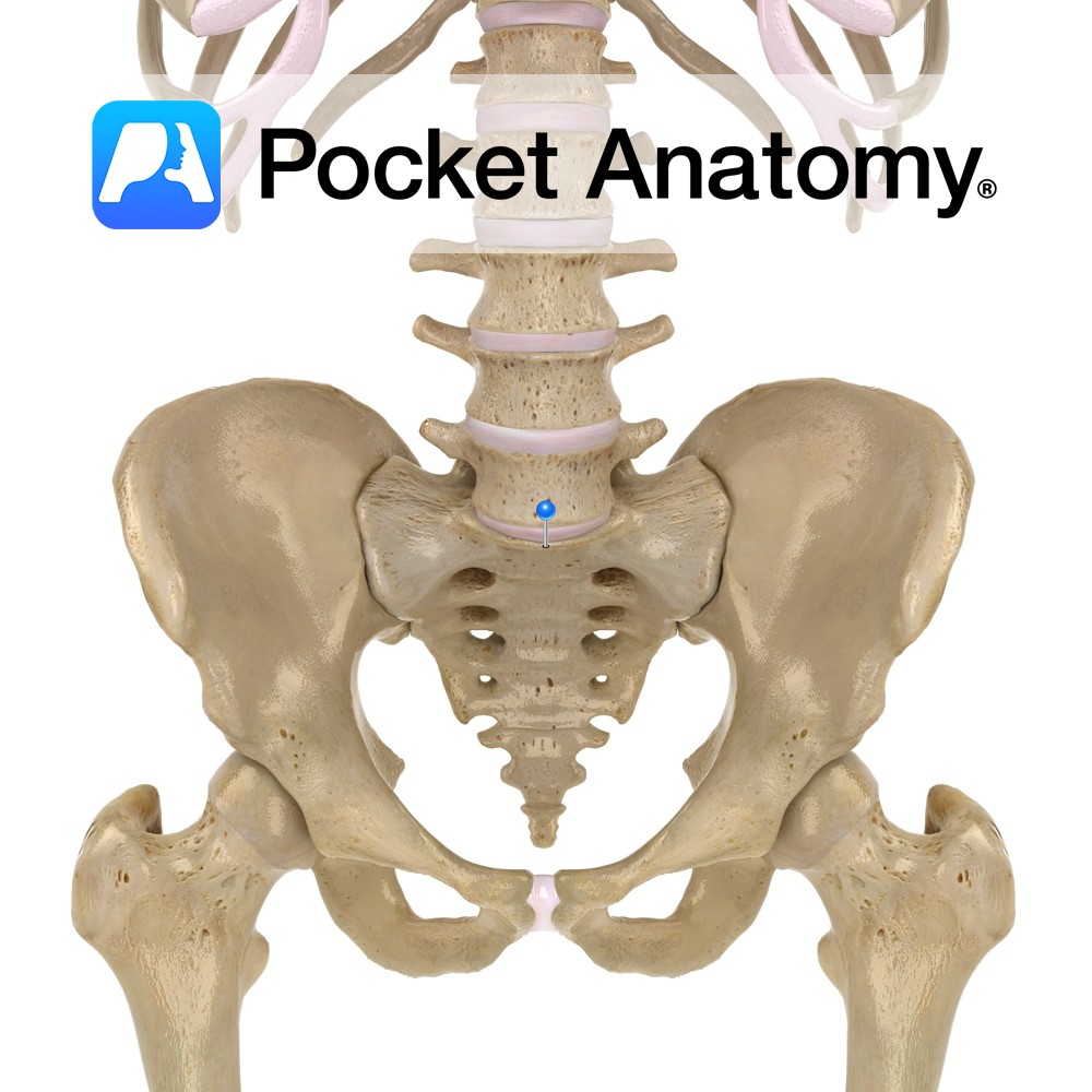
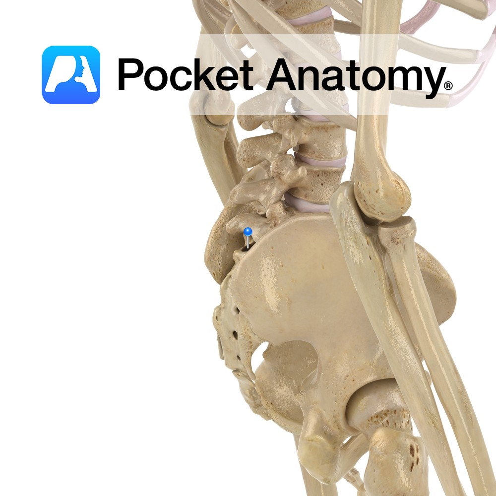
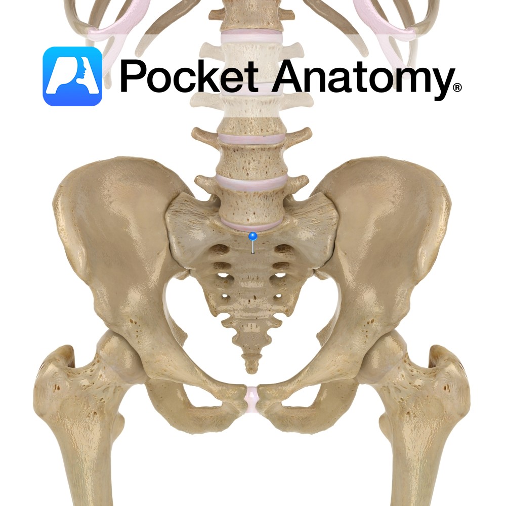
Anatomy Bulge forward where top (ie base) of wedge-shaped sacrum, where it joins with bottom (ie L5) of lumbar spine. Along with ileopectineal line, comprises the pelvic brim, edge of pelvic inlet (above, abdominal cavity; below, pelvic cavity). Sacrum tilts forward, presenting a sacrovertebral angle with lumbar spine; more prominent in female. Vignette Sacrum (Latin);
- Published in Pocket Anatomy Pins
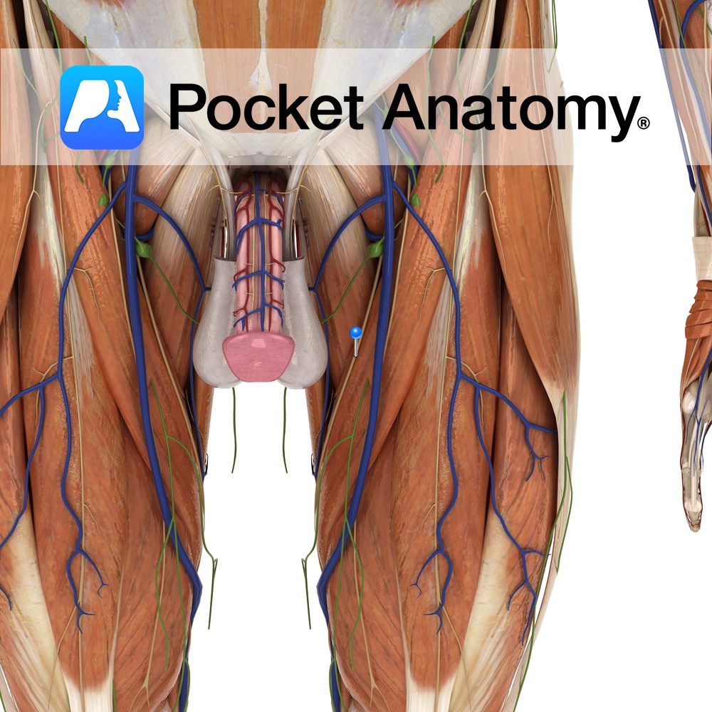
Anatomy Course A branch of the femoral nerve, arising after the femoral nerve passes under the inguinal ligament. It courses distally just behind the sartorius muscle. It does not travel through the adductor canal, but pierces through its aponeurotic covering to arise on the medial side of the knee, between the attachments of the sartorius
- Published in Pocket Anatomy Pins

Anatomy Origin: Anterior superior iliac spine of hip bone and superior part of notch below it. Insertion: Attaches by aponeurosis to medial surface of the tibia. Key Relations: -One of the five muscles of the anterior compartment of the thigh. -In the upper portion of the thigh sartorius forms the lateral margin of the femoral
- Published in Pocket Anatomy Pins
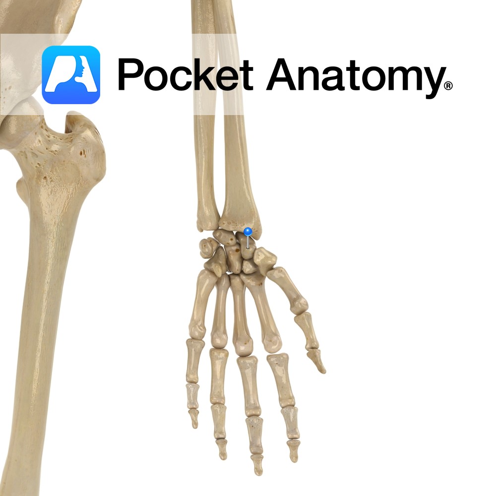
Anatomy Thumb side (radial) of nearer row of carpal bones, shape and size of cashew nut. Articulates up (proximally) with radius (lunate is other carpal that articulates with radius), down (distally) with trapezoid and trapezium, in (medially) with capitate and lunate. Clinical Commonest carpal bone fracture (60%). Requires prompt treatment. Pain, swelling and tenderness at
- Published in Pocket Anatomy Pins
Anatomy At birth, there are 5 (usually) distinct sacral bones; fusion starts age 16-18, finished about 34. S1 is the top (i.e. base) and biggest segment of the fused sacrum. Consists of broad, expanded large body in front of triangular sacral canal, foramina either side at bottom, ridge where fused with S2. Clinical Articulates with
- Published in Pocket Anatomy Pins
Anatomy At birth, 5 (usually) distinct sacral bones. Fusion starts age 16-18, finished about 34. S5 is lowermost (ie apex of inverted wedge shape) and smallest segment of the fused sacrum. Laminae don’t meet (leaving a sacral hiatus); no spinous process. Clinical Articulates with coccyx below. Interested in taking our award-winning Pocket Anatomy app for
- Published in Pocket Anatomy Pins
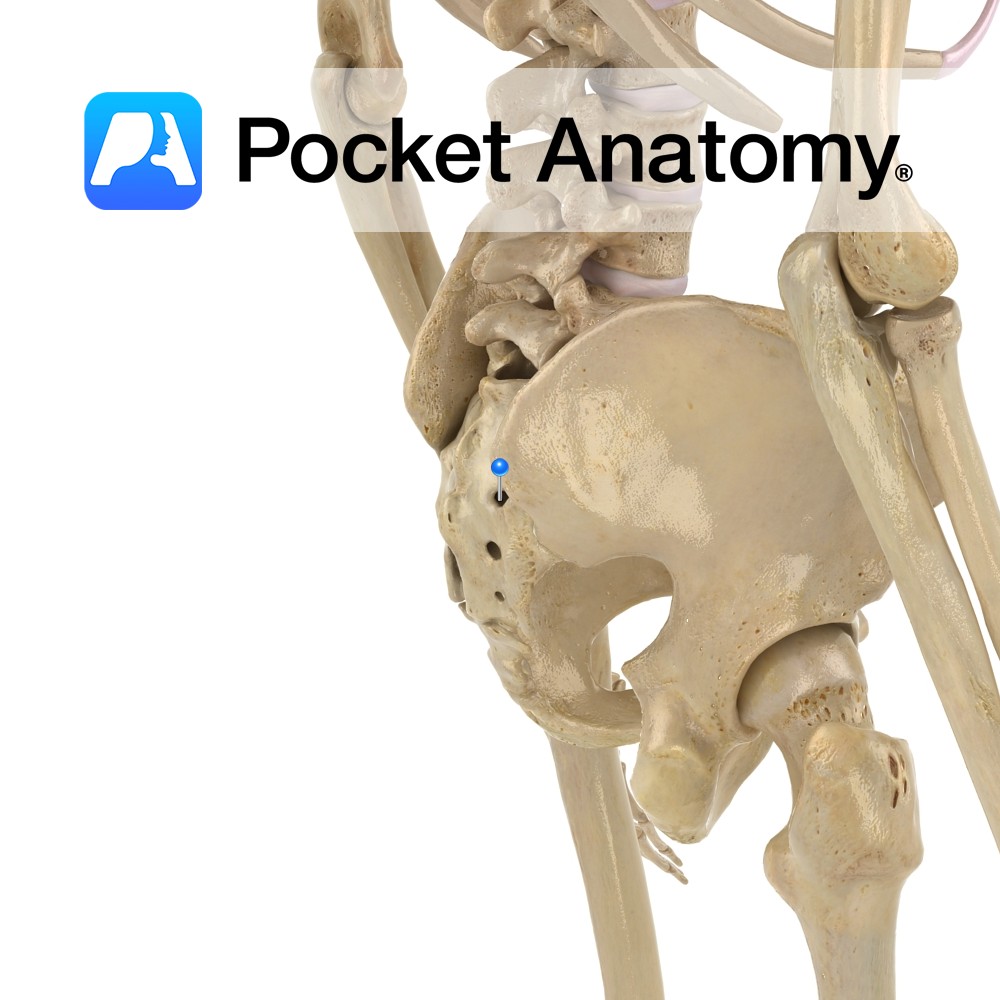
Anatomy Whereas cervical and thoracic vertebrae are distinct and afford passage of nerves through intervertebral foramina created by notches of adjacent pedicles, the fused sacrum retains foramina for this purpose, front and behind, at the lateral ends of ridges which are remnants (after fusion) of planes of separation of sacral vertebrae. Interested in taking our
- Published in Pocket Anatomy Pins
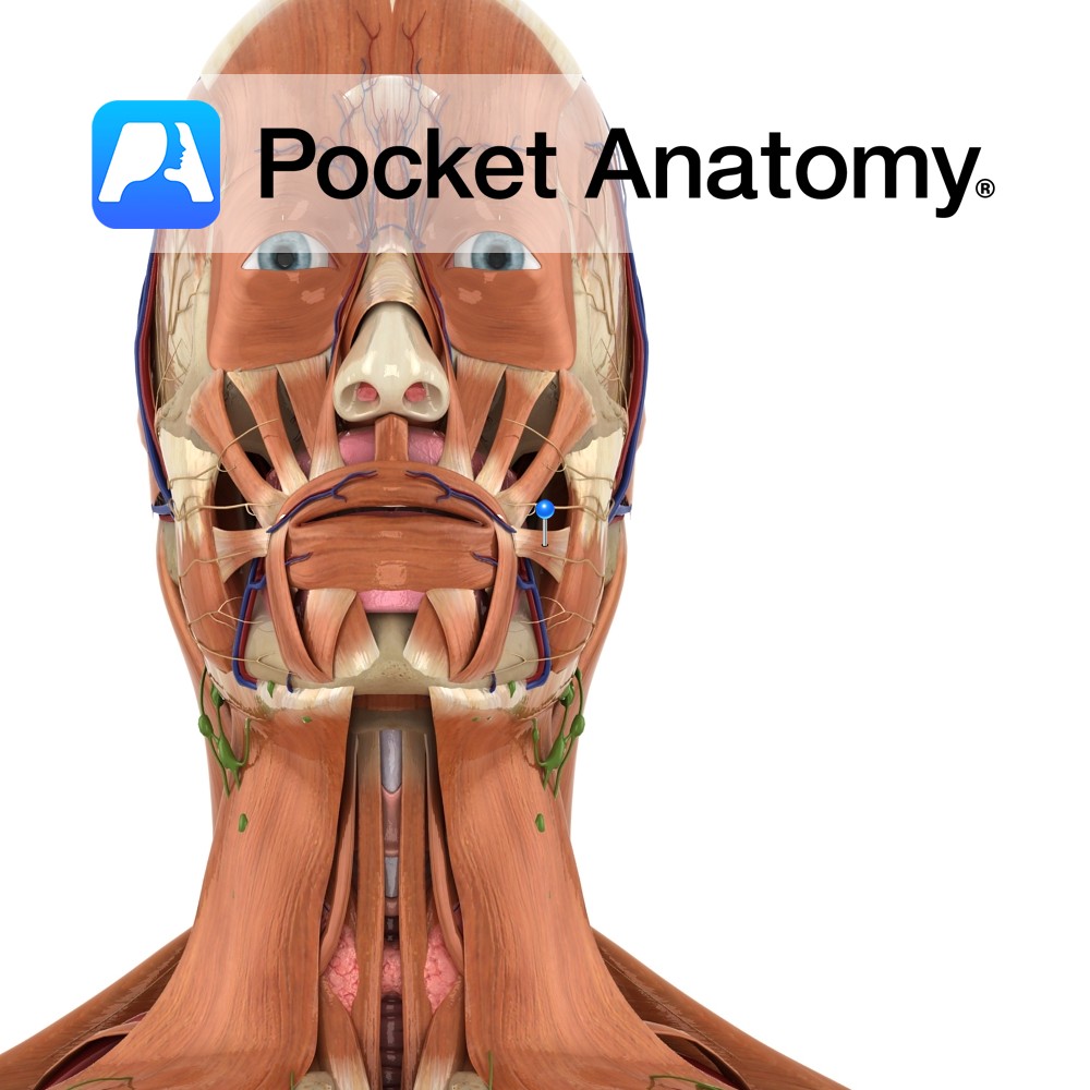
Anatomy Origin: Fascia over the masseter muscle. Insertion: Skin at the corner of the mouth. Key Relations: Thin superficial muscle in the upper group of oral muscles. Functions Pulls the corner of the mouth laterally and upwards. e.g. when grinning.. Supply Nerve Supply: Buccal branch of the facial nerve (CN 7). Blood Supply: Superior labial
- Published in Pocket Anatomy Pins