PocketAnatomy® is a registered brand name owned by © eMedia Interactive Ltd, 2009-2022.
iPhone, iPad, iPad Pro and Mac are trademarks of Apple Inc., registered in the U.S. and other countries. App Store is a service mark of Apple Inc.
Anatomy Origin: Ligamentum nuchae and spinous processes of C7 to T3 and supraspinous ligaments. Insertion: Upper border of 2nd to the 5th ribs just lateral to their angles. Key Relations: -Serratus posterior superior lies superior to the thoracolumbar fascia. -Sometimes referred to as part of the respiratory group. Functions -Elevates 2nd to the 5th ribs,
- Published in Pocket Anatomy Pins
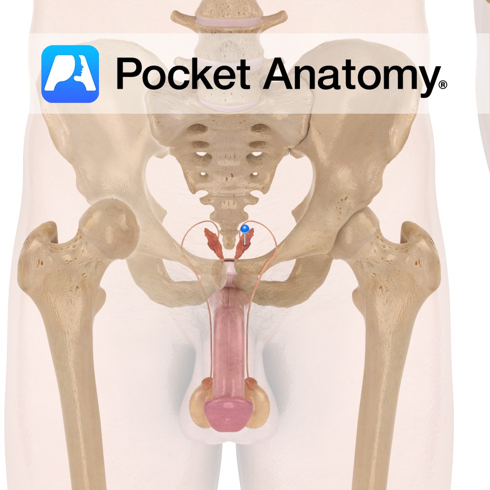
Functions Creates a significant portion (50-70%) of seminal fluid, though not all is released in the primary ejaculation (which consists mainly of prostatic fluid). The seminal vesicle empties into the vas deferens. (Note that the seminal vesicle forms in utero from a pouch of the vas deferens.). Anatomy Pair of tubular glands attached close to
- Published in Pocket Anatomy Pins

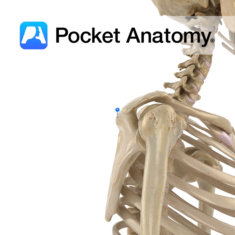
Anatomy Thickened axillary continuation (bony process) of spine of scapula, arching and extending laterally to overhang the gleno-humeral joint posteriorly, as the coracoid process does anteriorly. Articulates with the collarbone (clavicle). Clinical Highest point (summit) of shoulder. Its muscle and ligament attachments (as with the coracoid process) stabilize the shoulder joint, given the shallow socket
- Published in Pocket Anatomy Pins
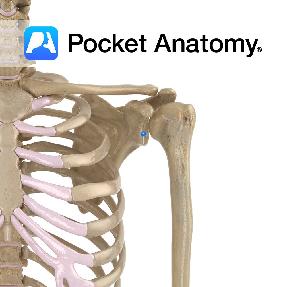
Anatomy Small hook-like bulge protruding laterally from top of axillary edge of scapula, over-hanging the gleno-humeral joint anteriorly, as the acromium does posteriorly. Clinical Its muscle and ligament attachments include the coracoclavicular ligament and (as with the acromium) they stabilize the shoulder joint, given the socket shallowness necessary to afford such a big range of
- Published in Pocket Anatomy Pins
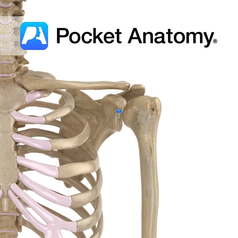
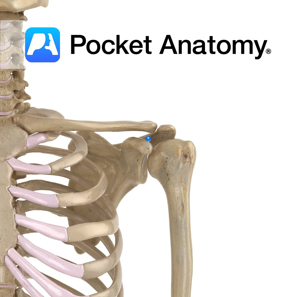
Anatomy Shallow saucer-shaped articular thickening of upper aspect (lateral angle) of lateral (axillary) surface, forming the shoulder joint by its articulation with the head of the humerus. Clinical The stability of the shoulder joint is mainly due to strength of rotator cuff (musculo-ligamentous girdle; supra- and infra-spinatus, subscapularis and teres minor muscles, and many ligaments),
- Published in Pocket Anatomy Pins
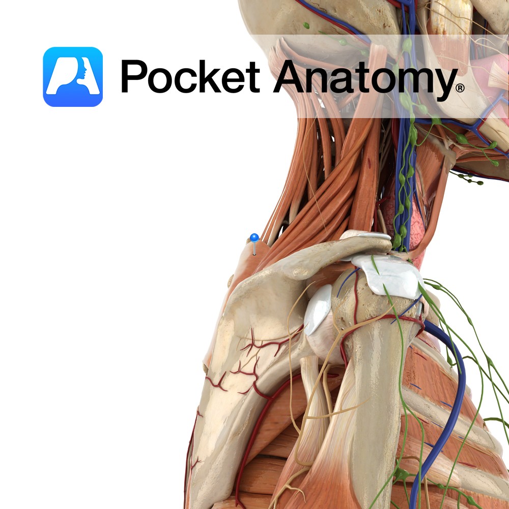
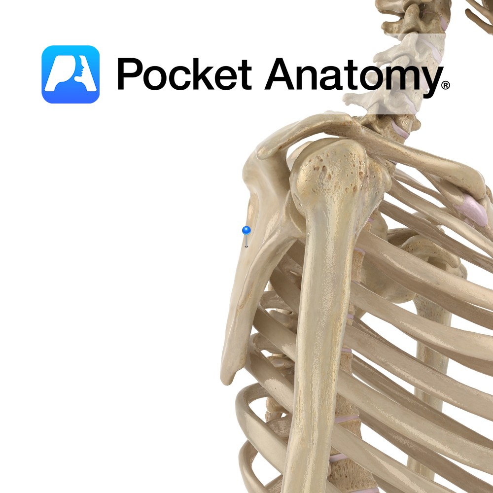
Anatomy Bulge just below the glenoid cavity (the socket of the ball-and-socket shoulder joint between scapula and humerus), on the lateral (axillary) border. Long head of triceps brachii attaches here. Clinical Triceps brachii (long head attached infraglenoid) extends the elbow, biceps brachii (long head attached supraglenoid) flexes the elbow and supinates the forearm (turns the
- Published in Pocket Anatomy Pins
Anatomy The back (dorsal, posterior) of the flat triangular part of the scapula, beneath the spine of the scapula which projects posteriorly above, making a fossa (there is a smaller supraspinous fossa above). Gives origin to infraspinatus (medial 2/3) and is covered by it (lateral 1/3). Vignette Fossa (Latin); ditch. Scapula fracture is ordinarily due
- Published in Pocket Anatomy Pins
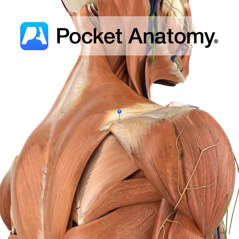
Anatomy The spine projects back from the dorsal part of the triangular scapula, about 3/4 way up, dividing the dorsal surface into small supraspinous fossa above and large infraspinous fossa below. Starting from flush with the medial (vertebral) border, it rises and thickens, becoming the acromium at its lateral (axillary) end/border. Clinical Many muscles attach;
- Published in Pocket Anatomy Pins
Anatomy The dorsal (posterior, back) of the flat triangular part of the scapula, above the spine of the scapula which projects posteriorly below, making a fossa (there is a bigger supraspinous fossa below. Gives origin to supraspinatus. Interested in taking our award-winning Pocket Anatomy app for a test drive?
- Published in Pocket Anatomy Pins
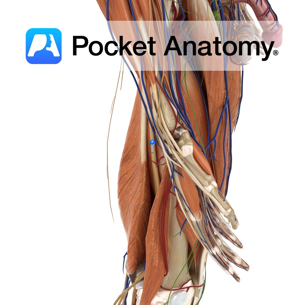
Anatomy Course The largest nerve in the body, arising from the lumbar plexus. It is made up of fibres from the anterior rami of L4 – S3. It originates at the posterior pelvic wall, and exits the pelvic cavity by passing through the greater sciatic foramen inferior to the piriformis muscle. It travels towards the
- Published in Pocket Anatomy Pins