PocketAnatomy® is a registered brand name owned by © eMedia Interactive Ltd, 2009-2022.
iPhone, iPad, iPad Pro and Mac are trademarks of Apple Inc., registered in the U.S. and other countries. App Store is a service mark of Apple Inc.

Anatomy Unpaired bone at base of skull, in front of temporal bones, behind eye socket. Looks like bat or butterfly from front. Made up of body, greater and lesser wings, and pterygoid processes. Body has sella tursica in which pituitary gland sits. Clinical Called “Keystone bone of cranium”, as it articulates with all other cranial
- Published in Pocket Anatomy Pins
Anatomy The dura mater of the spinal cord is the outermost layer of the meninges that surrounds the brain and spinal cord. It is the toughest of the three layers and surrounds the spinal cord like a loose fitting sock. Interested in taking our award-winning Pocket Anatomy app for a test drive?
- Published in Pocket Anatomy Pins
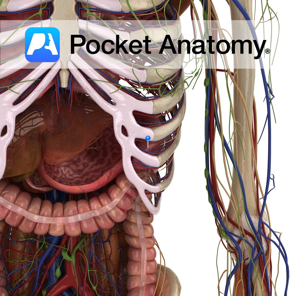
Anatomy Highly vascular, purplish, wedge-shaped (upper part thicker) organ (largest ductless), left upper quadrant abdomen; soft, as a result moulded by its contacts; diaphragmatic surface convex – diaphragm; visceral surface has anterior/ gastric, posterior/renal parts; gastric surface – back of fundus stomach, tail pancreas; renal surface – upper pole left kidney, sometimes adrenal; lower extremity
- Published in Pocket Anatomy Pins
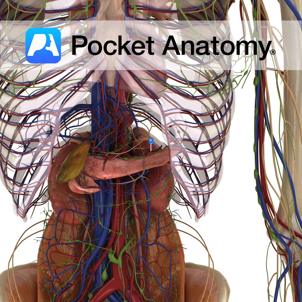
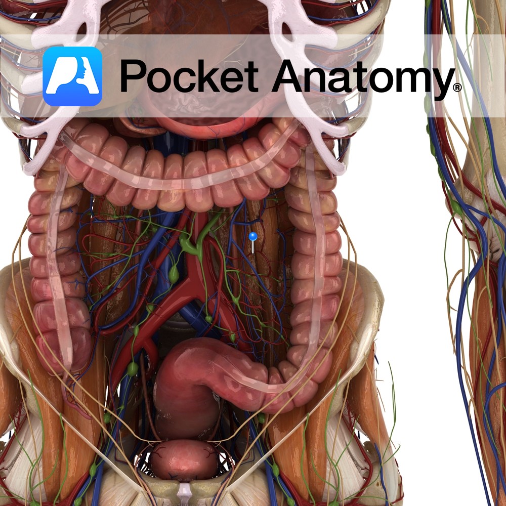
Anatomy Course Largest branch of the celiac trunk of the abdominal aorta. It travels laterally from the celiac trunk within the splenorenal ligament, taking a twisted course along the superior border of the pancreas, travelling posterior to the stomach. It divides into many small branches and enters the spleen at the hilum. Supply Supplies the
- Published in Pocket Anatomy Pins
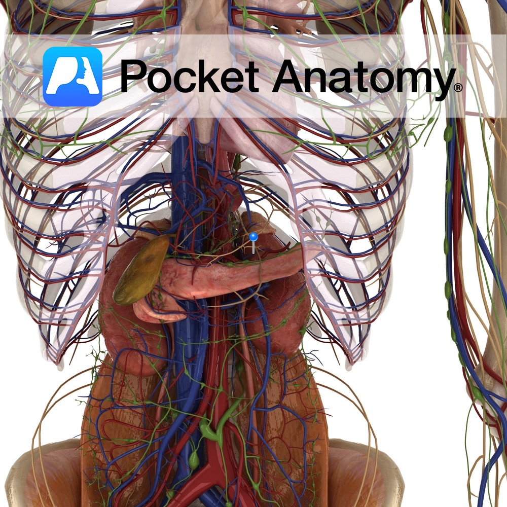
Anatomy Course Begins at the hilum of the spleen. It follows a tortuous course along the upper margin of the pancreas and travelling along the posterior abdominal wall, until it reaches the superior mesenteric vein, which it joins with to form the portal vein. Drain Drains blood from the spleen. Interested in taking our award-winning
- Published in Pocket Anatomy Pins
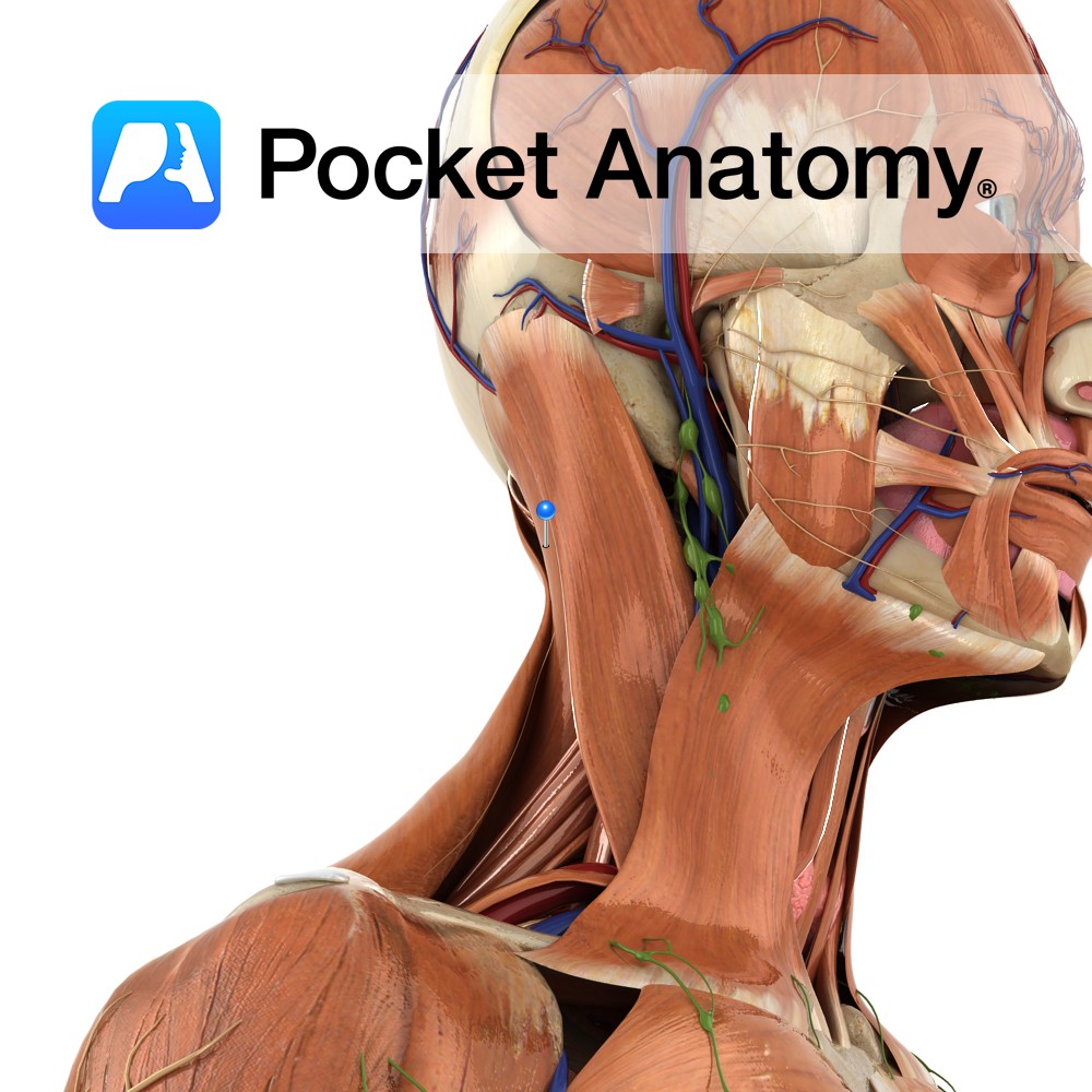
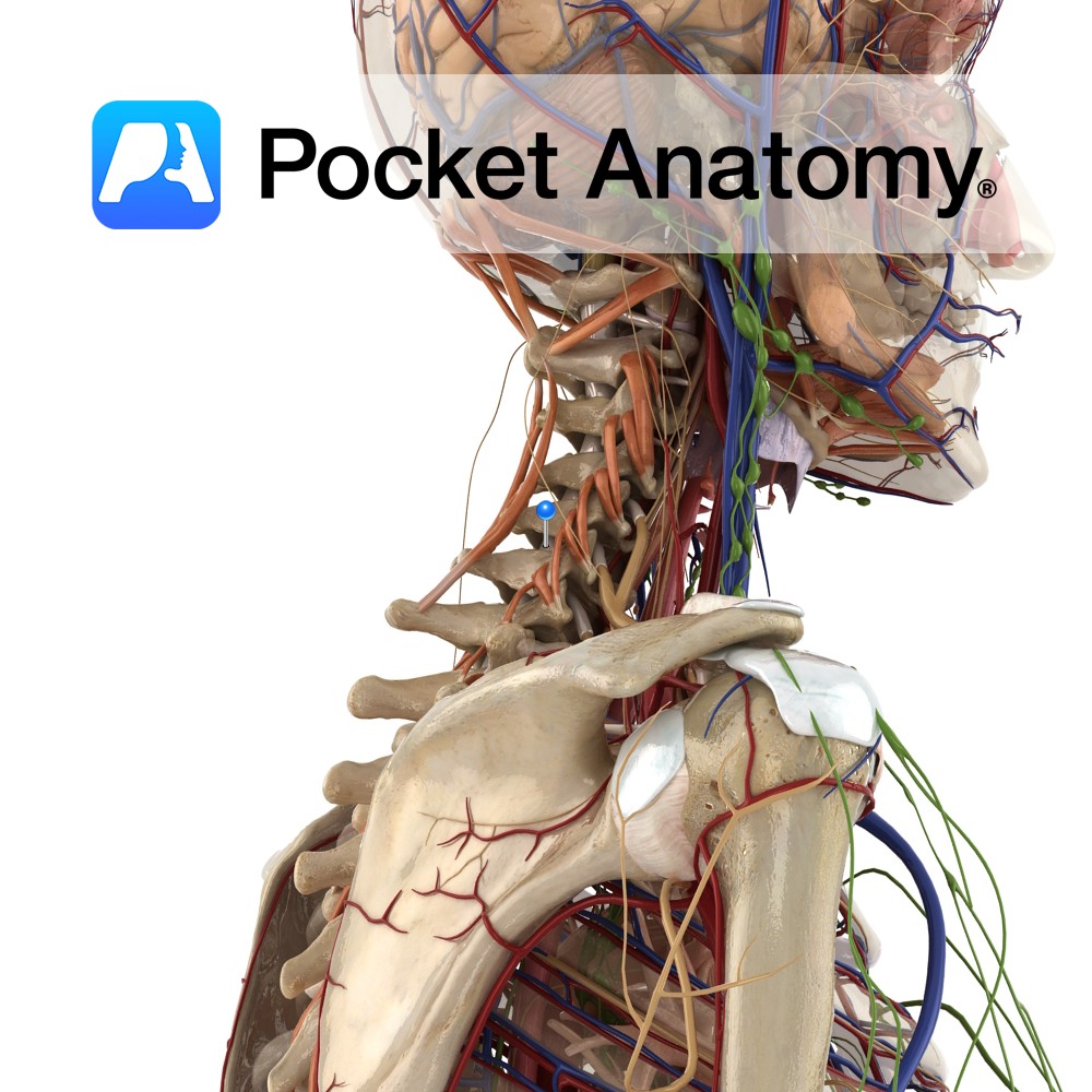
Anatomy Origin: Lower half of ligamentum nuchae and spinous processes of C7 to T4. Insertion: Mastoid process of the temporal bone and just below lateral third of the superior nuchal line on the occipital bone. Key Relations: -Superficial to spinalis capitis, semispinalis capitis and longissimus capitis and deep to sternocleidomasteoid. -Forms part of the floor
- Published in Pocket Anatomy Pins
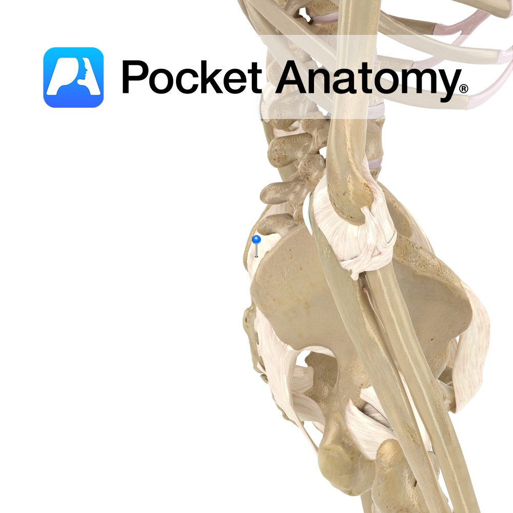
Anatomy Horizontal in direction, attaching from the first and second transverse tubercles on the posterior surface of the sacrum to the tuberosity of the ilium. Functions Supplies static stability to the sacroiliac joint. Interested in taking our award-winning Pocket Anatomy app for a test drive?
- Published in Pocket Anatomy Pins
.jpg)
Motion The glenohumeral joint is a multiaxial synovial ball and socket joint and involves the articulation between the glenoid fossa of the scapula and the head of the humerus. The glenohumeral joint is the most mobile joint in the body. It is capable of flexion, extension, abduction, adduction, medial rotation, lateral rotation, and circumduction. Circumduction
- Published in Pocket Anatomy Pins
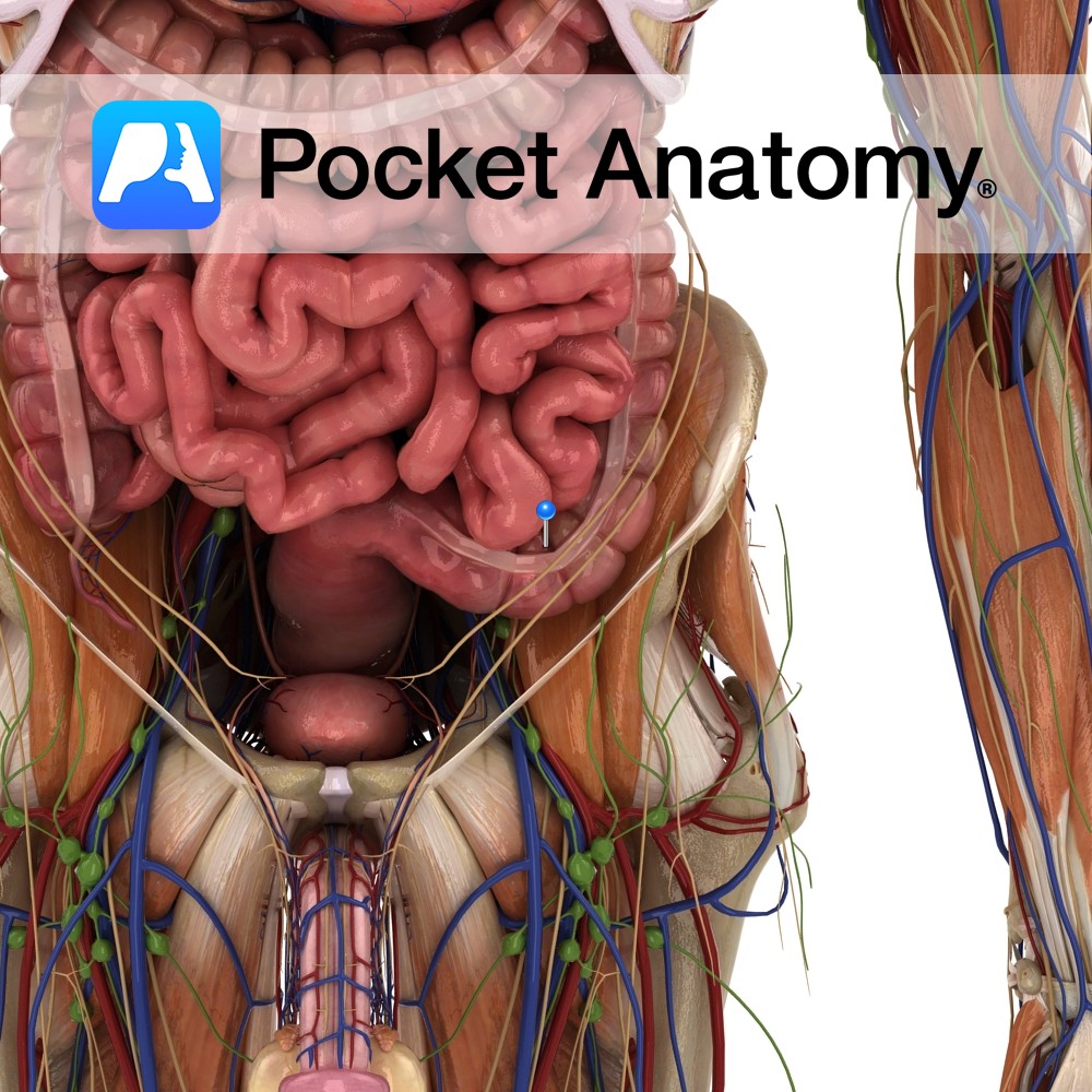
Anatomy Course There can be two to four sigmoid arteries. These are branches from the inferior mesenteric artery that descend towards the sigmoid colon in the lower left quadrant of the abdomen. They travel within the sigmoid mesocolon and anastomose with branches from the left colic artery and the superior rectal artery. Supply Supply sigmoid
- Published in Pocket Anatomy Pins
Anatomy Loop of colon continuous above with descending colon, ordinarily within pelvis (mobile, can be displaced), starting at pelvic brim, passing across in front of sacrum from left to right, then back left to midline at level S3 (3rd fused sacral vertebra), then down to be continuous with rectum, surrounded by peritoneum with a mesocolon,
- Published in Pocket Anatomy Pins