PocketAnatomy® is a registered brand name owned by © eMedia Interactive Ltd, 2009-2022.
iPhone, iPad, iPad Pro and Mac are trademarks of Apple Inc., registered in the U.S. and other countries. App Store is a service mark of Apple Inc.
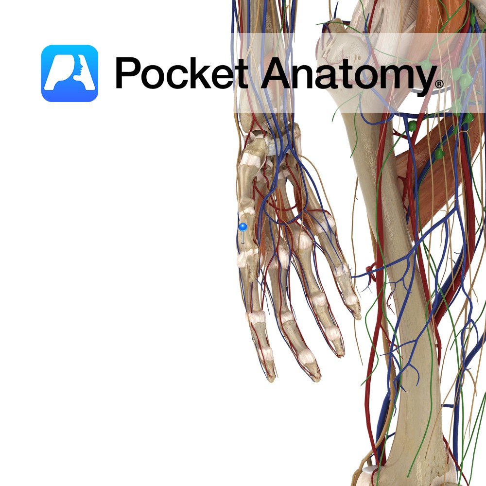
Anatomy Origin: Middle thirds of posterior of radius and ulna and intervening interosseus membrane. Insertion: Lateral aspect of base of 1st metacarpal. Key Relations: -Its tendon together with the tendon of extensor pollicis brevis forms the anterior boundary of the anatomical snuff box. -One of the six muscles in the deep posterior compartment of the
- Published in Pocket Anatomy Pins
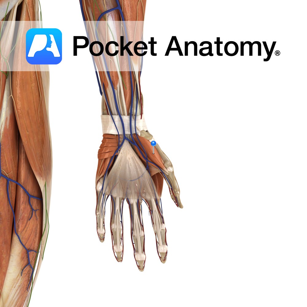
Anatomy Origin: Tubercles of scaphoid and trapezium and adjacent flexor retinaculum. Insertion: Proximal phalanx and extensor apparatus of the thumb. Key Relations: Is one of the muscles of the thenar eminence of the hand. Functions -Abduction of the thumb at the carpopmetacarpol and metacarpophalangeal joints. -Also assists in thumb opposition and extension. Supply Nerve Supply:
- Published in Pocket Anatomy Pins
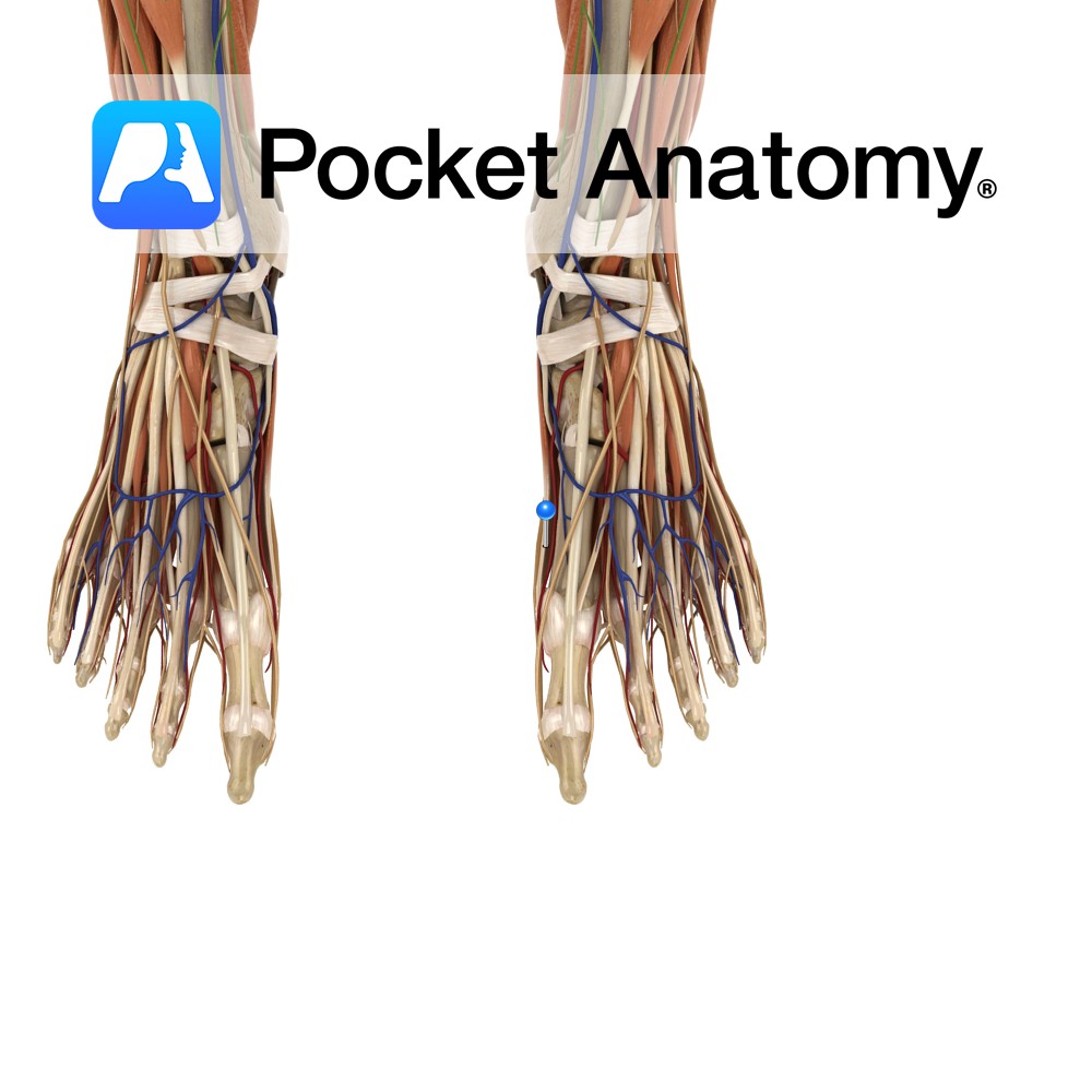
Anatomy Origin: Medial process of the calcaneal tuberosity, flexor retinaculum and plantar aponeurosis. Insertion: Medial surface of the base of the proximal phalanx of the hallux (big toe). Key relations: Lies on the medial surface of the foot and contributes to a soft tissue bulge on the medial part of the sole of the foot.
- Published in Pocket Anatomy Pins
Anatomy Origin: Pisiform and tendon of flexor carpi ulnaris. Insertion: Ulnar aspect of the base of the proximal phalanx of the little finger and ulnar border of the extensor apparatus of the little finger. Key Relations: Is one of the muscles of the hypothenar eminence of the hand. Functions Abduction of the little finger at
- Published in Pocket Anatomy Pins
Anatomy Origin: Lateral and medial processes of the calcaneal tuberosity and plantar aponeurosis. Insertion: Lateral surface of the base of the 5th proximal phalanx. Key relations: -Lies on the lateral surface of the foot. -The lateral plantar vessels and nerve located medially. Functions Abducts the 5th toe at the metatarsophalangeal joint. Supply Nerve supply: Lateral
- Published in Pocket Anatomy Pins
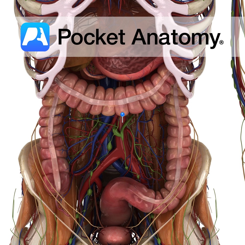
Anatomy Course Enters the abdomen as a continuation of the thoracic aorta by passing through the aortic hiatus of the diaphragm at the level of T12. Descends downward slightly to the left of the midline until it bifurcates into the left and right common iliac arteries at the level to L4. The inferior vena cava
- Published in Pocket Anatomy Pins
After the completion of the Skeletal System, Connective Tissues and Muscular System, the next phase was to construct the internal organs of the reproductive, digestive, and respiratory systems.
- Published in 3D Human, Teaching Anatomy
After the completion of the Skeletal System and Connective Tissues, the next phase was to construct the muscular system, which enables the body to move, maintain posture, and circulate blood and consists of skeletal, smooth and cardiac muscles. Firstly the creation of the muscular system was approached in very much the same way as the
- Published in 3D Human, Teaching Anatomy
After completion of the skeletal system, building the cartilage, ligaments, and connective tissues within the body was the next logical phase as along with the bones, these are the fibrous connective tissues that also provide structural support and protection within the body. All of the connective tissue was sculpted by hand in ZBrush on top of the skeleton model. After
- Published in 3D Human, Teaching Anatomy
In June 2016, our team started on a journey to develop a 3D Human Anatomy Model. As the skeleton is the support structure of the human body – giving the body its shape and form, providing attachment points for muscles to allow movement of the joints and serving as a frame for protecting the organs
- Published in 3D Human


.jpg)
.jpg)


