PocketAnatomy® is a registered brand name owned by © eMedia Interactive Ltd, 2009-2022.
iPhone, iPad, iPad Pro and Mac are trademarks of Apple Inc., registered in the U.S. and other countries. App Store is a service mark of Apple Inc.
Anatomy Articulates up with middle phalanx at 5th DIP. Interested in taking our award-winning Pocket Anatomy app for a test drive?
- Published in Pocket Anatomy Pins
Anatomy Articulates up with middle phalanx at 3rd DIP. Interested in taking our award-winning Pocket Anatomy app for a test drive?
- Published in Pocket Anatomy Pins
Anatomy The more distal of the 2 thumb phalanges, connected above by the intermediate interphalangeal joint. Interested in taking our award-winning Pocket Anatomy app for a test drive?
- Published in Pocket Anatomy Pins
Anatomy Articulates up with 5th middle phalanx. Interested in taking our award-winning Pocket Anatomy app for a test drive?
- Published in Pocket Anatomy Pins
Anatomy Articulates up with 3rd middle phalanx. Interested in taking our award-winning Pocket Anatomy app for a test drive?
- Published in Pocket Anatomy Pins
Anatomy Articulates proximally with 1st proximal phalanx. Interested in taking our award-winning Pocket Anatomy app for a test drive?
- Published in Pocket Anatomy Pins
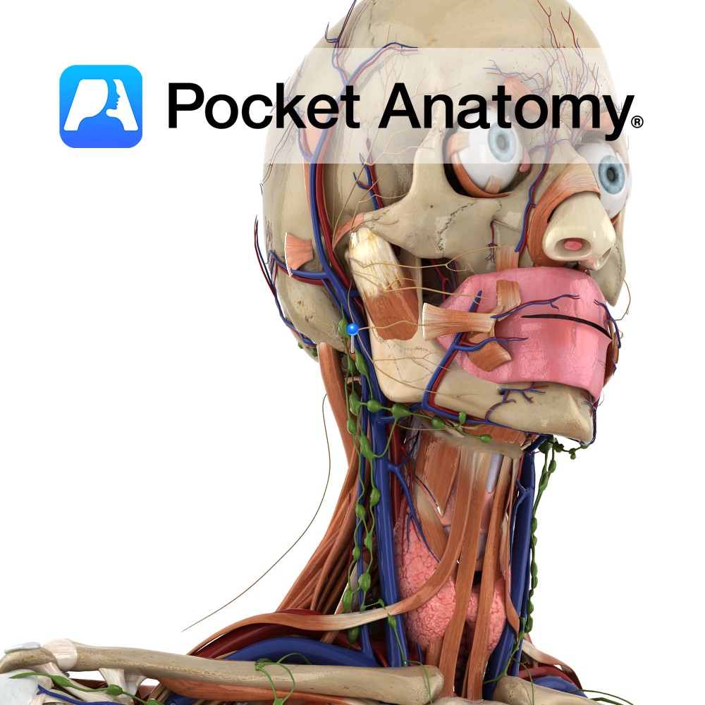
Anatomy Origin: Mastoid notch on medial side of mastoid process of temporal bone. Insertion: Intermediate tendon between the two bellies which itself attaches to the body of the hyoid bone. Key Relations: -Is one of the suprahyoid muscles lying in the anterior triangle of the neck. -Forms inferoposterior boundary of submandibular triangle. -Forms superior boundary
- Published in Pocket Anatomy Pins
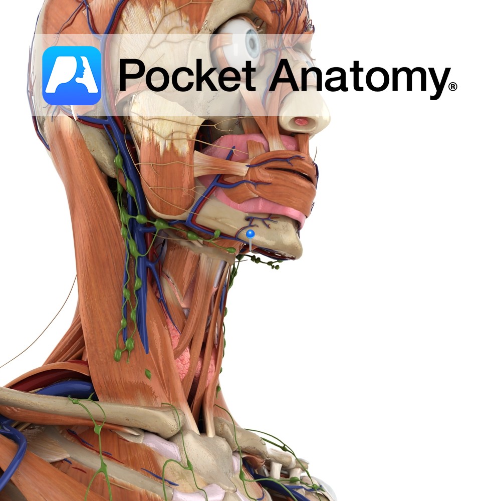
Anatomy Origin: Digastric fossa on inside of mandible. Insertion: Intermediate tendon between the two bellies which itself attaches to the body of the hyoid bone. Key Relations: -Is one of the suprahyoid muscles lying in the anterior triangle of the neck. -Forms inferoanterior boundary of submandibular triangle. -Forms lateral boundary of submental triangle. -Superior to
- Published in Pocket Anatomy Pins
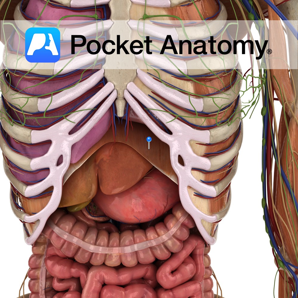
Anatomy Origin: Xiphoid process, costal margin, lower six costal cartilages, ends of 11th and 12th ribs and lumbar vertebrae L1 to L3. Insertion: Central tendon of diaphragm. Key relations: -The inferior vena cava passes through the caval hiatus of the diaphragm at vertebral level T8. -The oesophagus and vagus nerve pass through the oesophageal hiatus
- Published in Pocket Anatomy Pins
Anatomy Course Branch of the genicular artery that pierces the adductor canal, where it then travels to the medial aspect of the knee with the saphenous nerve. Supply Anastomotic network around the knee. Interested in taking our award-winning Pocket Anatomy app for a test drive?
- Published in Pocket Anatomy Pins

.jpg)
.jpg)
.jpg)
.jpg)
.jpg)
.jpg)


.jpg)