PocketAnatomy® is a registered brand name owned by © eMedia Interactive Ltd, 2009-2022.
iPhone, iPad, iPad Pro and Mac are trademarks of Apple Inc., registered in the U.S. and other countries. App Store is a service mark of Apple Inc.
Anatomy Course Arises at the bifurcation of the common iliac artery where it then descends to the upper aspect of the greater sciatic foramen. Supply Responsible for supplying the pelvic wall and floor, pelvic viscera as well as the erectile tissue of the perineum. Interested in taking our award-winning Pocket Anatomy app for a test
- Published in Pocket Anatomy Pins
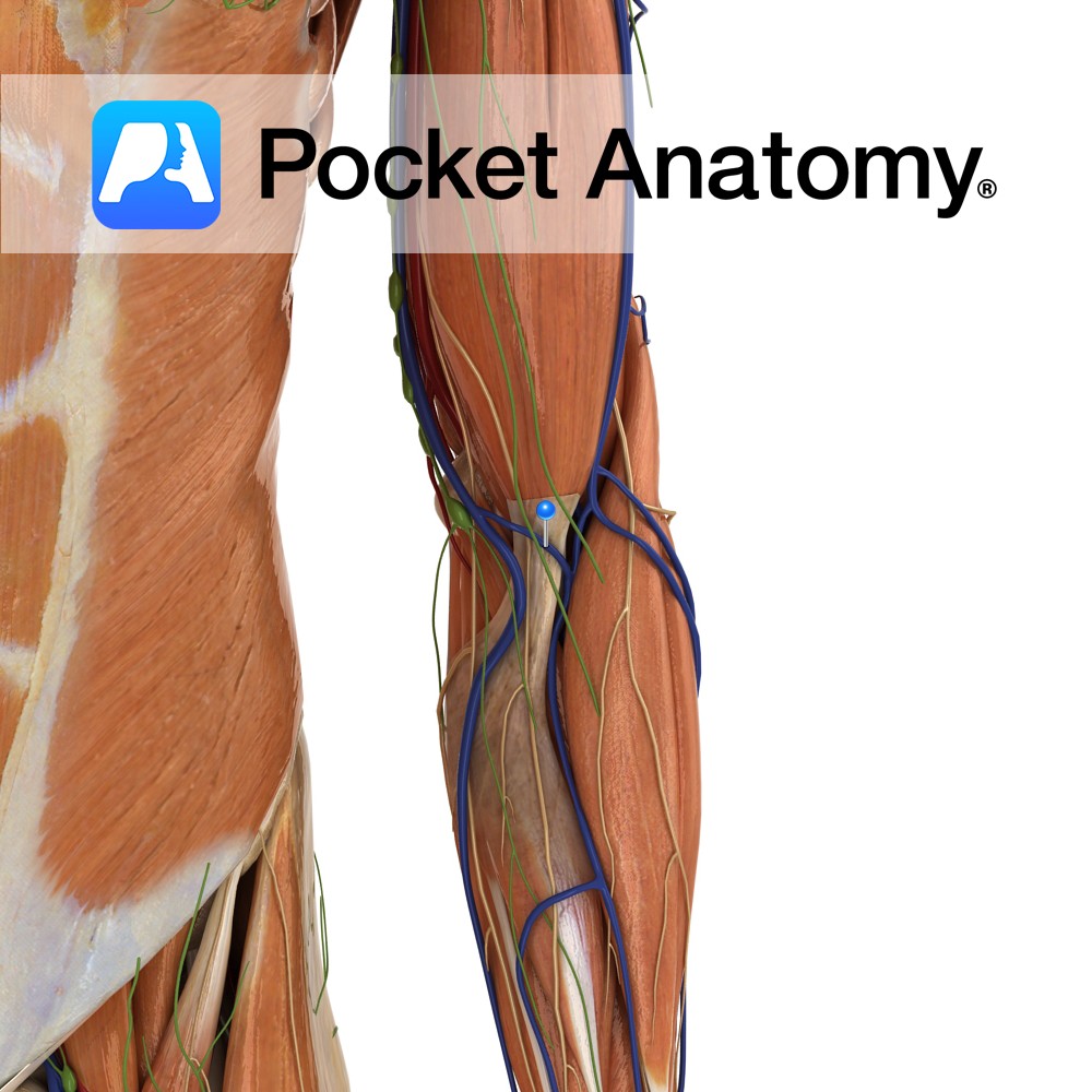
Anatomy Course One of several superficial veins that join the cephalic and basilic vein in a “H” like manner. Drain Drains the forearm. Interested in taking our award-winning Pocket Anatomy app for a test drive?
- Published in Pocket Anatomy Pins
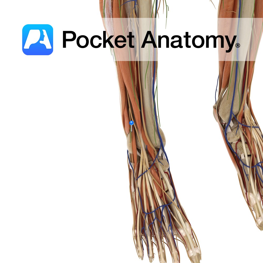
Anatomy Course The terminal branch of superficial peroneal nerve. Travels on the lateral side of the foot where it eventually gives off dorsal digital branches. Supply Supplies the skin of the dorsum digits of the foot with the exception of the lateral aspect of the little toe and in between the great and second toe.
- Published in Pocket Anatomy Pins
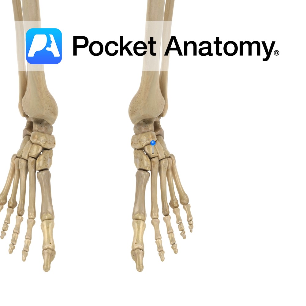
Anatomy Tarsal bone, wedge-shaped, smallest of the cuneiforms, between lateral and medial cuneiforms, articulates forward with 2nd metatarsal, with cuneiforms either side, back with navicular. Vignette Ankle (talocrural) joint; tibia and fibula with tarsal. Subtalar joint; talar with calcaneus. Transverse Talar joint; talonavicular and calcaneocuboid. Tarsometatarsal joints (arthrodial/sliding); 3 cuneiforms and cuboid with metatarsals. Interested
- Published in Pocket Anatomy Pins
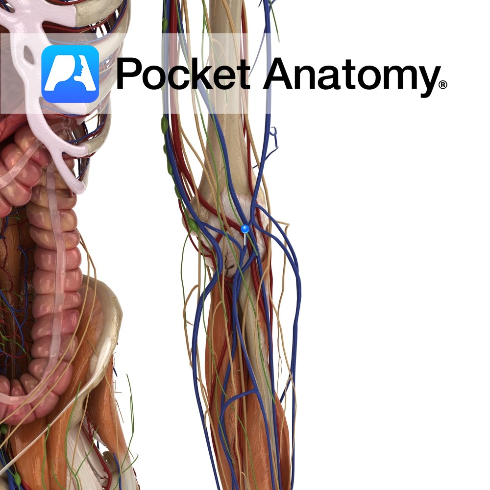
Anatomy Course A medium sized vein that comes off the antebrachial vein. At the elbow joint it joins the cephalic vein. Drain Drains the superficial component of the forearm. Clinical It is located significantly farther from arteries than other veins. Along with its size, it makes a popular target for inserting a cannula. Interested in
- Published in Pocket Anatomy Pins
Anatomy Course Anterior division of the ninth thoracic spinal nerve. Runs along the ninth intercostal space, below the superior rib until it ends as the anterior cutaneous branch which surfaces to supply the cutaneous skin. Along the way it gives off a lateral cutaneous branch that emerges superficially and splits into a posterior and anterior
- Published in Pocket Anatomy Pins
Anatomy Course Anterior division of the eighth thoracic spinal nerve. Runs along the eighth intercostal space, below the superior rib until it ends as the anterior cutaneous branch which surfaces to supply the cutaneous skin. Along the way it gives off a lateral cutaneous branch that emerges superficially and splits into a posterior and anterior
- Published in Pocket Anatomy Pins
Anatomy Course Anterior division of the seventh thoracic spinal nerve. Runs along the seventh intercostal space, below the superior rib until it ends as the anterior cutaneous branch which surfaces to supply the cutaneous skin. Along the way it gives off a lateral cutaneous branch that emerges superficially and splits into a posterior and anterior
- Published in Pocket Anatomy Pins
Anatomy Course Anterior division of the sixth thoracic spinal nerve. Runs along the sixth intercostal space, below the superior rib until it ends as the anterior cutaneous branch which surfaces to supply the cutaneous skin. Along the way it gives off a lateral cutaneous branch that emerges superficially and splits into a posterior and anterior
- Published in Pocket Anatomy Pins
Anatomy Course Anterior division of the fifth thoracic spinal nerve. Runs along the fifth intercostal space, below the superior rib until it ends as the anterior cutaneous branch which surfaces to supply the cutaneous skin. Along the way it gives off a lateral cutaneous branch that emerges superficially and splits into a posterior and anterior
- Published in Pocket Anatomy Pins

