PocketAnatomy® is a registered brand name owned by © eMedia Interactive Ltd, 2009-2022.
iPhone, iPad, iPad Pro and Mac are trademarks of Apple Inc., registered in the U.S. and other countries. App Store is a service mark of Apple Inc.
Anatomy Origin: Middle thirds of posterior of radius and ulna and intervening interosseus membrane. Insertion: Lateral aspect of base of 1st metacarpal. Key Relations: -Its tendon together with the tendon of extensor pollicis brevis forms the anterior boundary of the anatomical snuff box. -One of the six muscles in the deep posterior compartment of the
- Published in Pocket Anatomy Pins
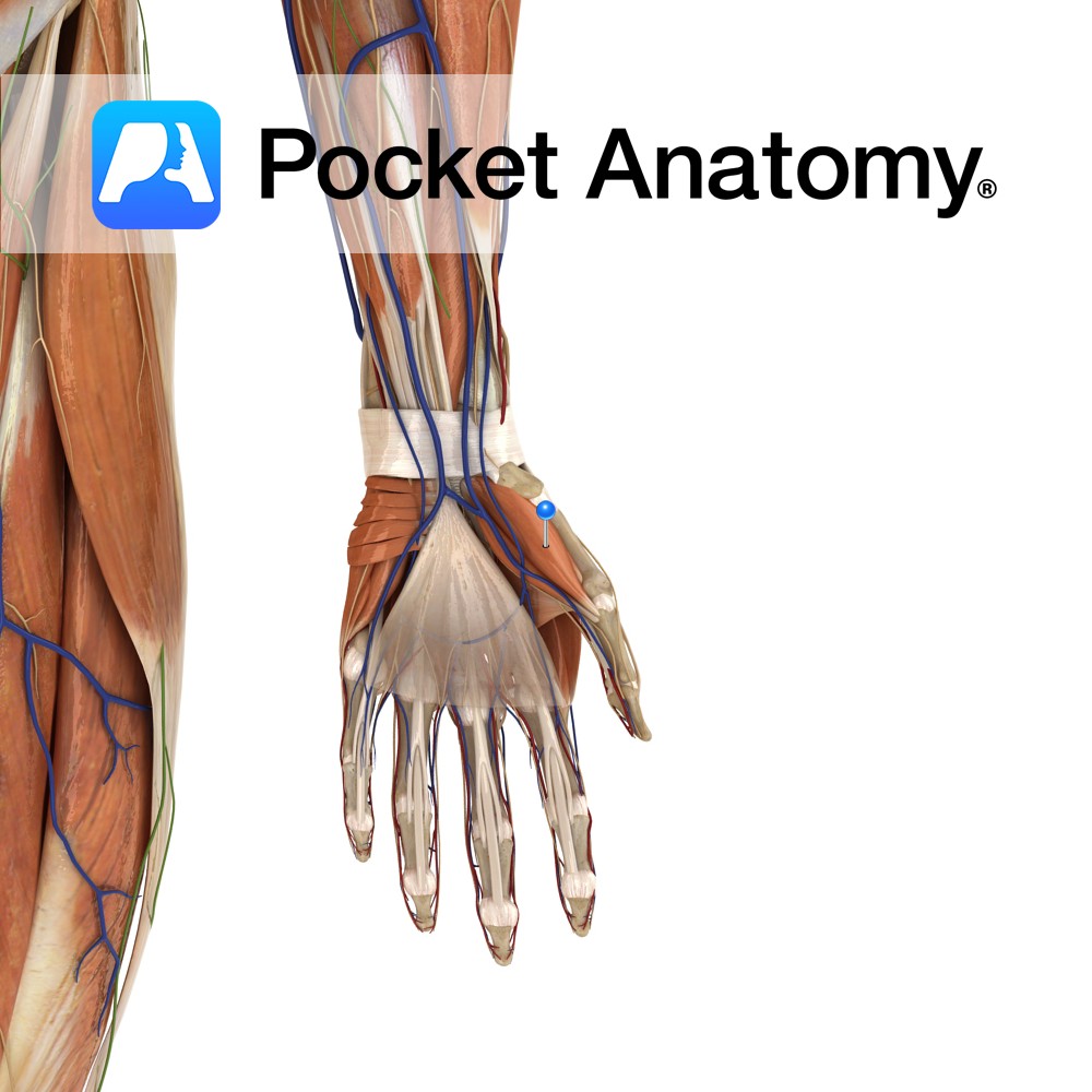
Anatomy Origin: Tubercles of scaphoid and trapezium and adjacent flexor retinaculum. Insertion: Proximal phalanx and extensor apparatus of the thumb. Key Relations: Is one of the muscles of the thenar eminence of the hand. Functions -Abduction of the thumb at the carpopmetacarpol and metacarpophalangeal joints. -Also assists in thumb opposition and extension. Supply Nerve Supply:
- Published in Pocket Anatomy Pins
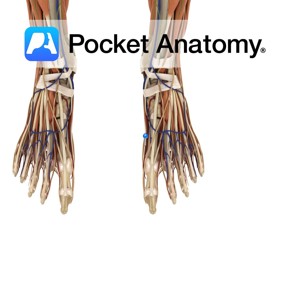
Anatomy Origin: Medial process of the calcaneal tuberosity, flexor retinaculum and plantar aponeurosis. Insertion: Medial surface of the base of the proximal phalanx of the hallux (big toe). Key relations: Lies on the medial surface of the foot and contributes to a soft tissue bulge on the medial part of the sole of the foot.
- Published in Pocket Anatomy Pins
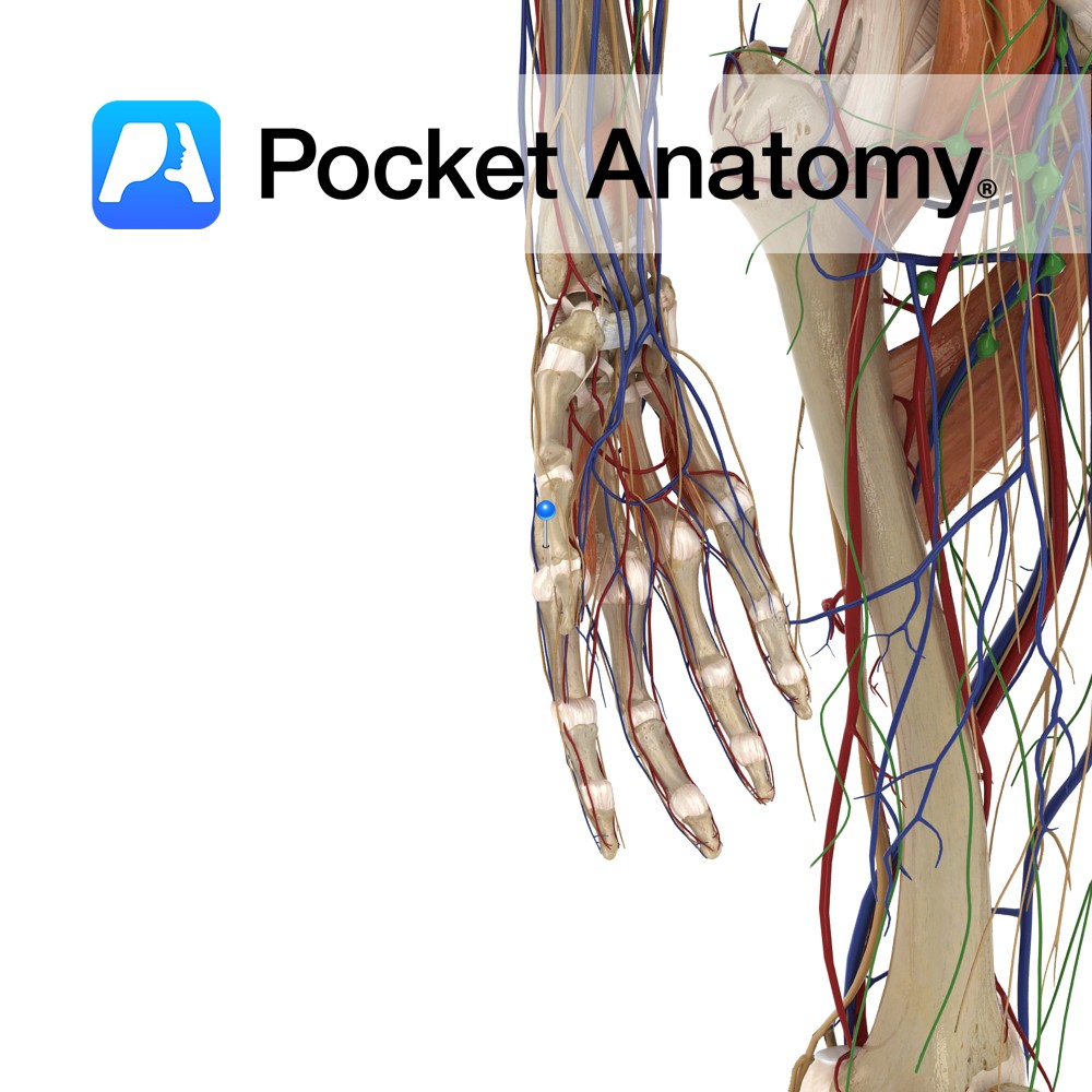
Anatomy Origin: Pisiform and tendon of flexor carpi ulnaris. Insertion: Ulnar aspect of the base of the proximal phalanx of the little finger and ulnar border of the extensor apparatus of the little finger. Key Relations: Is one of the muscles of the hypothenar eminence of the hand. Functions Abduction of the little finger at
- Published in Pocket Anatomy Pins
Anatomy Origin: Lateral and medial processes of the calcaneal tuberosity and plantar aponeurosis. Insertion: Lateral surface of the base of the 5th proximal phalanx. Key relations: -Lies on the lateral surface of the foot. -The lateral plantar vessels and nerve located medially. Functions Abducts the 5th toe at the metatarsophalangeal joint. Supply Nerve supply: Lateral
- Published in Pocket Anatomy Pins
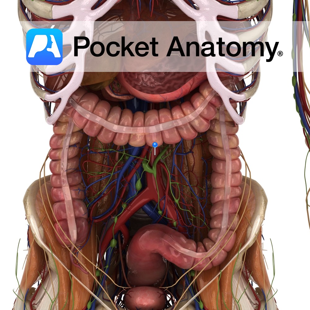
Anatomy Course Enters the abdomen as a continuation of the thoracic aorta by passing through the aortic hiatus of the diaphragm at the level of T12. Descends downward slightly to the left of the midline until it bifurcates into the left and right common iliac arteries at the level to L4. The inferior vena cava
- Published in Pocket Anatomy Pins
Pocket Anatomy has won the “Boost” StartUp competition at The Next Web (TNW) Conference in Amsterdam. The event is one of Europe’s top tech gatherings and was attended by over 2,500 influential web, technology and business leaders from all over the world. Nearly a hundred StartUps were selected for Boost, out of which 10 (including
- Published in Awards
To celebrate the launch of the new iOS7 operating system update on iPad and iPhone, Pocket Anatomy announces its beautiful and minimalist new interface! <H2>New and Improved</H2> Harnessing the already highly user-friendly 3D interface and features of previous versions, the newest refresh of Pocket Anatomy is a vital addition to anyone’s library of medical Apps.
- Published in App Updates
May 30th sees the launch of one of the most significant medical anatomy apps available on the Apple Platform. The 4th Generation update to the multi award-winning Pocket Anatomy now includes full female and male anatomy. This complements the recent additions of circulatory & lymphatic systems, as well as the inclusion of both cranial &
- Published in App Updates
Full text version: Users can navigate eight layers of brain anatomy, view cross sections and nerve pathways, insert notes, and peruse clinical findings. Elizabeth Armstrong Moore by Elizabeth Armstrong Moore March 26, 2012 1:09 PM PDT Of all the subjects best taught in 3D, anatomy has got to be up there. And when it comes
- Published in In the News

.jpg)
.jpg)
