PocketAnatomy® is a registered brand name owned by © eMedia Interactive Ltd, 2009-2022.
iPhone, iPad, iPad Pro and Mac are trademarks of Apple Inc., registered in the U.S. and other countries. App Store is a service mark of Apple Inc.
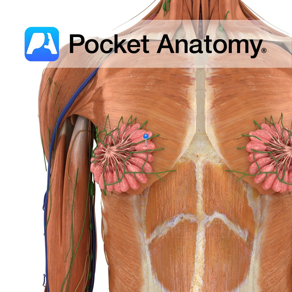
Anatomy Paired skin-covered mammary (milk-producing) gland (modified apocrine/sweat gland) ventral (in front of) pectoralis major muscle on thoracic/chest wall, levels ribs #2 to 6; can extend to axilla at side (sometimes as far as latissimus dorsi at back), up to clavicle, in/medial to sternum, made of adipose/glandular/connective/myoepithelial tissue, comprising 15-20 irregular lobes (each with 10-100
- Published in Pocket Anatomy Pins
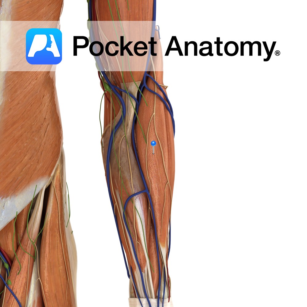
Anatomy Origin: Proximal part of the lateral supracondylar ridge of the humerus and adjacent intermuscular septum. Insertion: Lateral surface of the distal end of the radius. Key Relations: -One of the four muscles of the superficial posterior compartment of the forearm. -Lies anterior to the elbow joint. -Forms the lateral boundary of the cubital fossa.
- Published in Pocket Anatomy Pins
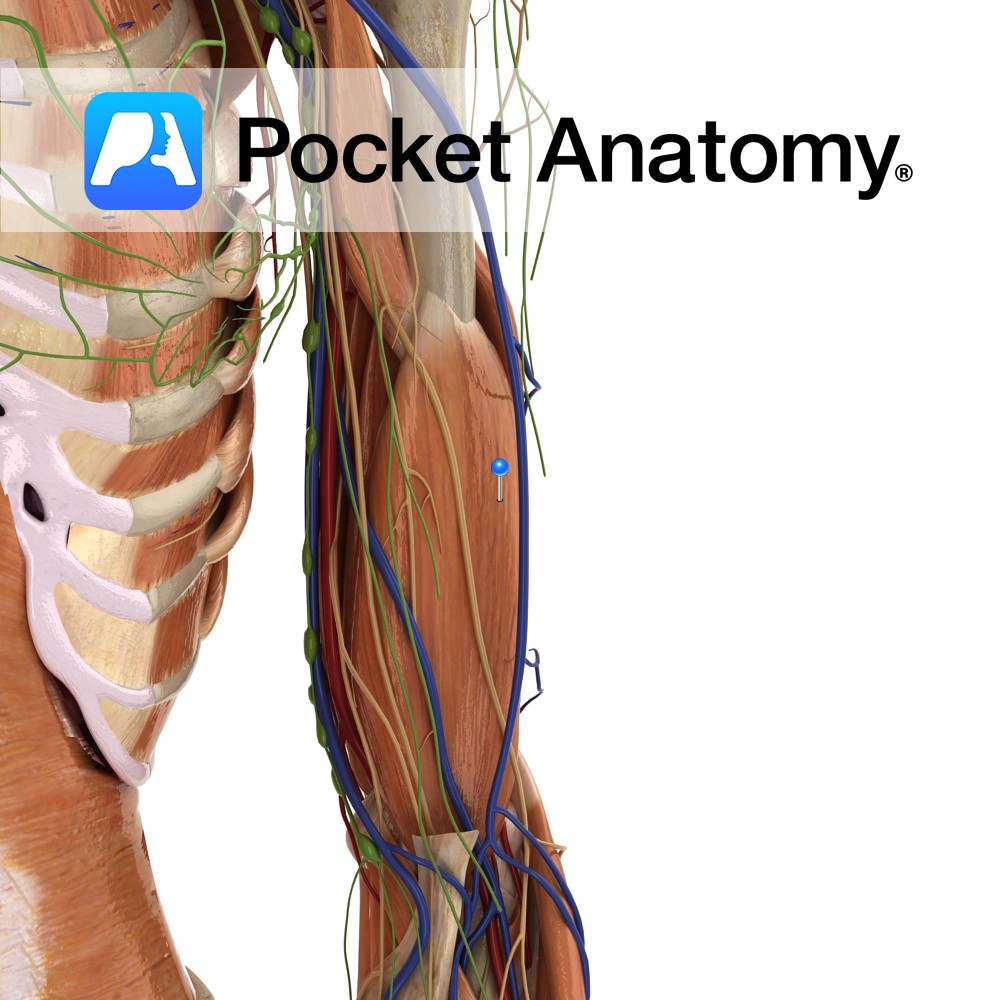
Anatomy Origin: Distal half of anterior surface of the humerus. Insertion: Coronoid process and the tuberosity of the ulna. Key Relations: -Located deep to the biceps brachii and forms part of the floor of the cubital fossa. –Musculocutaneous nerve lies on its lateral surface adjacent to biceps brachii. Functions Works synergistically with biceps brachii for
- Published in Pocket Anatomy Pins
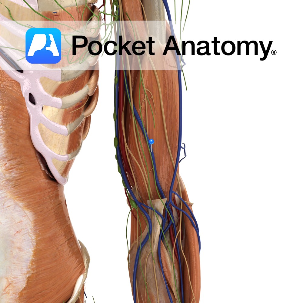
Anatomy Course Begins with the fusion of the radial and ulnar veins at the elbow. It then travels beneath the muscles of the forearm as it travels proximally up the arm. It eventually joins the basilic vein. Drain Drains the muscles of the forearm. Interested in taking our award-winning Pocket Anatomy app for a test
- Published in Pocket Anatomy Pins
Anatomy Course A network of nerve fibers running from the spine to neck, axilla and forearm. It is divided into roots (C5, C6, C7, C8, T1), trunks (superior, middle and inferior), divisions (3 anterior and 3 posterior), cords (lateral, posterior and medial), and terminal branches (musculocutaneous, axillary, radial, median and ulnar nerves). Supply Responsible for
- Published in Pocket Anatomy Pins


.jpg)
.jpg)
.jpg)
.jpg)
.jpg)
.jpg)