PocketAnatomy® is a registered brand name owned by © eMedia Interactive Ltd, 2009-2022.
iPhone, iPad, iPad Pro and Mac are trademarks of Apple Inc., registered in the U.S. and other countries. App Store is a service mark of Apple Inc.
Anatomy Course Arises from the lateral aspect of the axillary artery. Travels behind the coracobrachialis muscles and the short head of the biceps brachii and anterior to the neck of the humerus. Anastamoses with the posterior circumflex humeral artery. Supply Supplies the glenohumeral joint and the neck of the humerus. Interested in taking our award-winning
- Published in Pocket Anatomy Pins
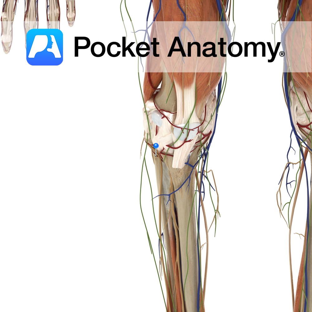
Anatomy Course Branch of the anterior tibial artery. Travels laterally through the soleus muscle as well as continuing around the neck of the fibula. It ends by contributing to the anastomotic network of the knee. Supply Supplies the soleus muscle and other adjacent muscles. Interested in taking our award-winning Pocket Anatomy app for a test
- Published in Pocket Anatomy Pins
Anatomy Pedicles project posteriorly from either side of the back of the vertebral body, joining laminae which themselves meet at the midline (from where a spinous process projects back and down) to complete the neural arch (enclosing the vertebral foramen). Where pedicle meets lamina each side, a transverse process, to which muscles and ligaments attach,
- Published in Pocket Anatomy Pins
Anatomy Pedicles project posteriorly from either side of the back of the vertebral body, joining laminae which themselves meet at the midline to complete the neural arch (enclosing the vertebral foramen). A spinous process projects back and down from this junction; muscles and ligaments attach to it. Interested in taking our award-winning Pocket Anatomy app
- Published in Pocket Anatomy Pins
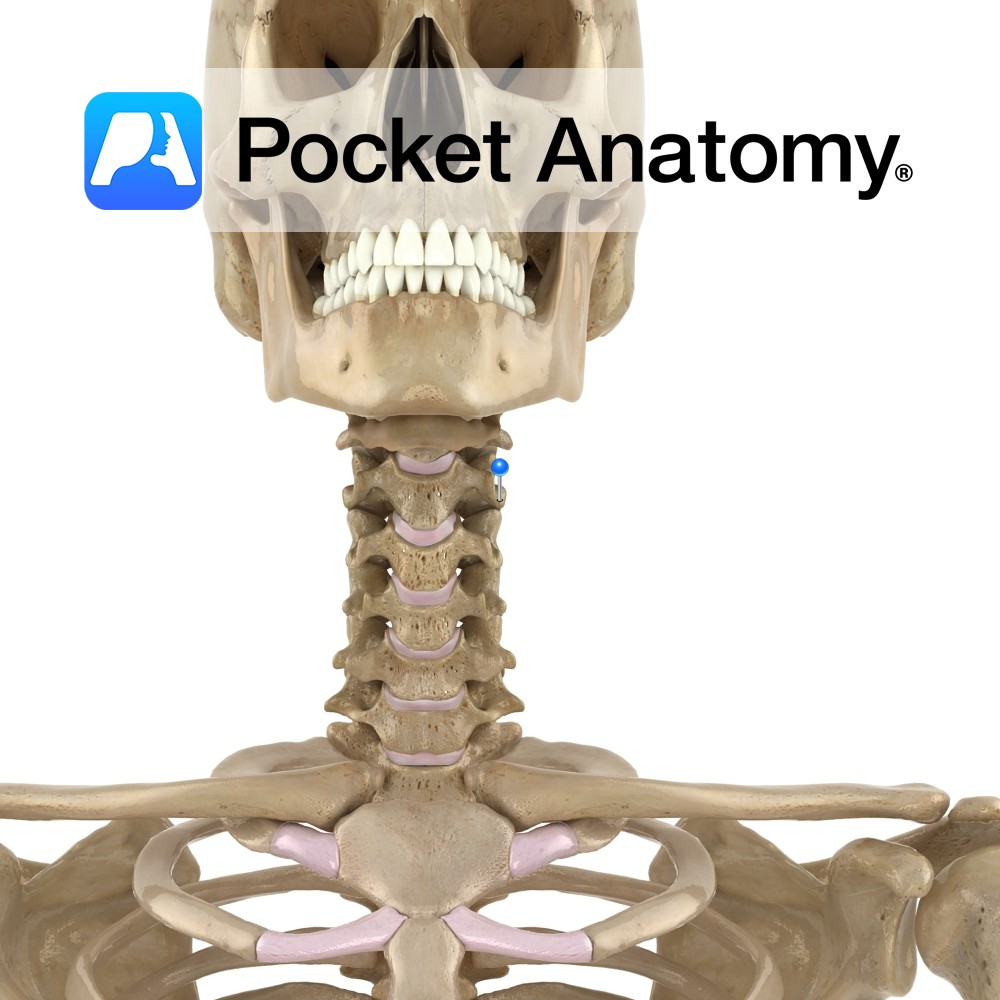
Anatomy Pedicles project posteriorly from either side of the back of the vertebral body, joining laminae which themselves meet at the midline (from where a spinous process projects back and down) to complete the neural arch (enclosing the vertebral foramen). Adjacent laminae above and below are connected by the ligamenta flava. Interested in taking our
- Published in Pocket Anatomy Pins
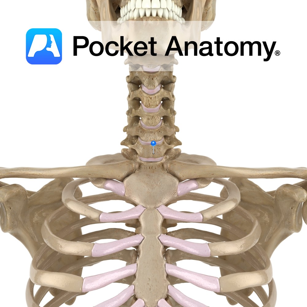
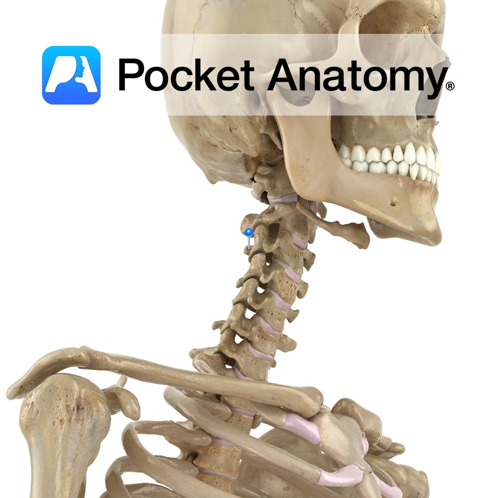
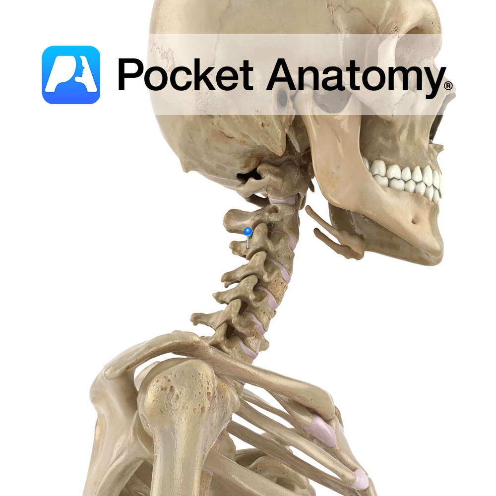
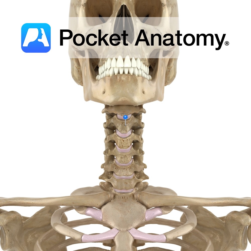
Anatomy Cylindrical, broader side-to-side, upper surface lipped at sides, lower surface indented at sides. Articulates with vertebrae above and below through fibrocartilaginous discs that allow some vertebral movement and provide shock absorption. No articular facets (unlike those on thoracic for articulation with ribs). Clinical The cervical (and lumbar) spine exhibits flexion, extension and some lateral
- Published in Pocket Anatomy Pins
Anatomy Most noticeable feature – odontoid process (derived from separation and fusion of body C1 with that of C2) rising from upper surface. Also large strong spinous process. C2 has complex articulation with atlas: Pivotal junction with odontoid process at 2 joints – front of odontoid with anterior arch C1, back of odontoid with transverse
- Published in Pocket Anatomy Pins
Anatomy Highest vertebra of column of 33. Forms spine/skull joint, along with C2 (axis). C1 has no vertebral body, as it is fused with that of C2 to contribute to formation of odontoid process (or dens). C1 has no spinous process; its absence affords greater range of movement of skull on C1. Superior facets (atlanto-occipital)
- Published in Pocket Anatomy Pins
Anatomy Also called vertbra prominens. Its spinous process is thick, long, almost horizontal, ends in a tubercle to which the bottom of the ligamentum nuchae is attached. In 70% of people, this is the most prominent spinous process (30% either C6 or T1). In 1 in 500, the transverse process has a forward projection as
- Published in Pocket Anatomy Pins
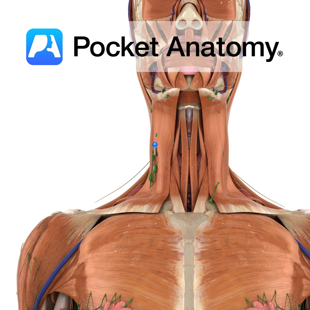
Anatomy Group/Cluster of nodes, in front and back of ears and along lower border jaw, and deeper in neck alongside blood vessels. Nodes filter the lymphatic drainage of scalp, face, nasal cavity, pharynx. Clinical Main (6) groups/clusters of nodes; Cervical, Axillary, Thoracic (Mediastinal, Bronchopulmonary), Iliac, Inguinal. Interested in taking our award-winning Pocket Anatomy app for
- Published in Pocket Anatomy Pins

.jpg)
.jpg)
.jpg)
