PocketAnatomy® is a registered brand name owned by © eMedia Interactive Ltd, 2009-2022.
iPhone, iPad, iPad Pro and Mac are trademarks of Apple Inc., registered in the U.S. and other countries. App Store is a service mark of Apple Inc.
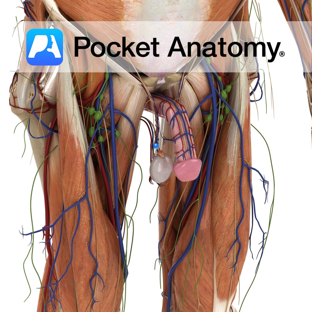
Anatomy 15-20 muscular ducts which connect rete testis to epididymis, accounting for 1/3 volume head of epididymis, concentrating spermatozoa by water reabsorption and moving them on. Interested in taking our award-winning Pocket Anatomy app for a test drive?
- Published in Pocket Anatomy Pins
Anatomy The dura mater is the strong, thick, outermost layer of the meninges attached to the periosteum of the skull except where it runs long the skull base or is reflected along the skull vault. Venous sinuses run inside spaces within the dura mater created by separation of the dura mater from the periosteum at
- Published in Pocket Anatomy Pins
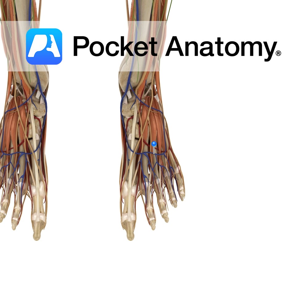
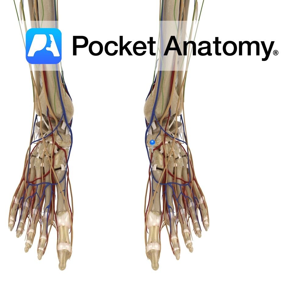
Anatomy Course A superficial vein that joins the great and small saphenous veins. It starts where the first dorsal digital vein of the foot meets with the great saphenous vein and connects to the small saphenous vein where it meets the fifth dorsal digital vein of the foot. Drain Receives tributaries from the dorsal digits
- Published in Pocket Anatomy Pins
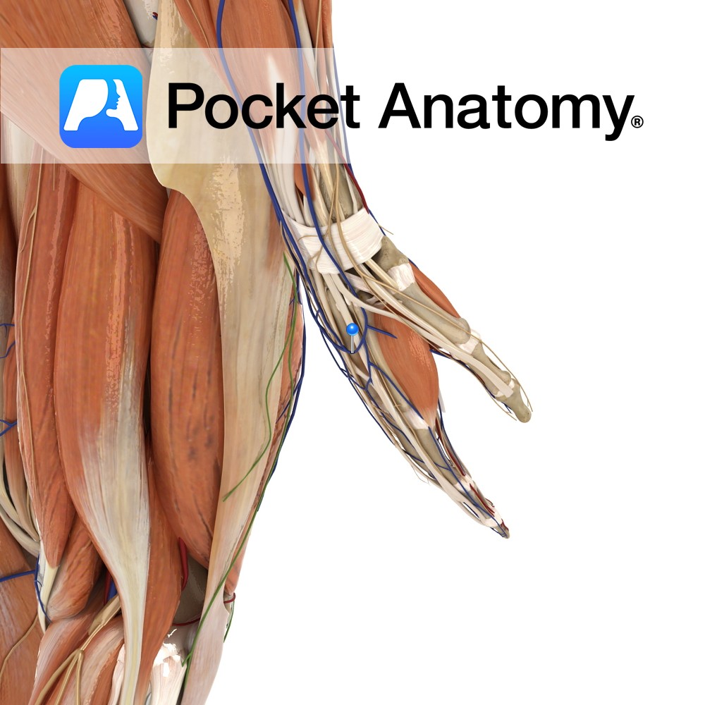
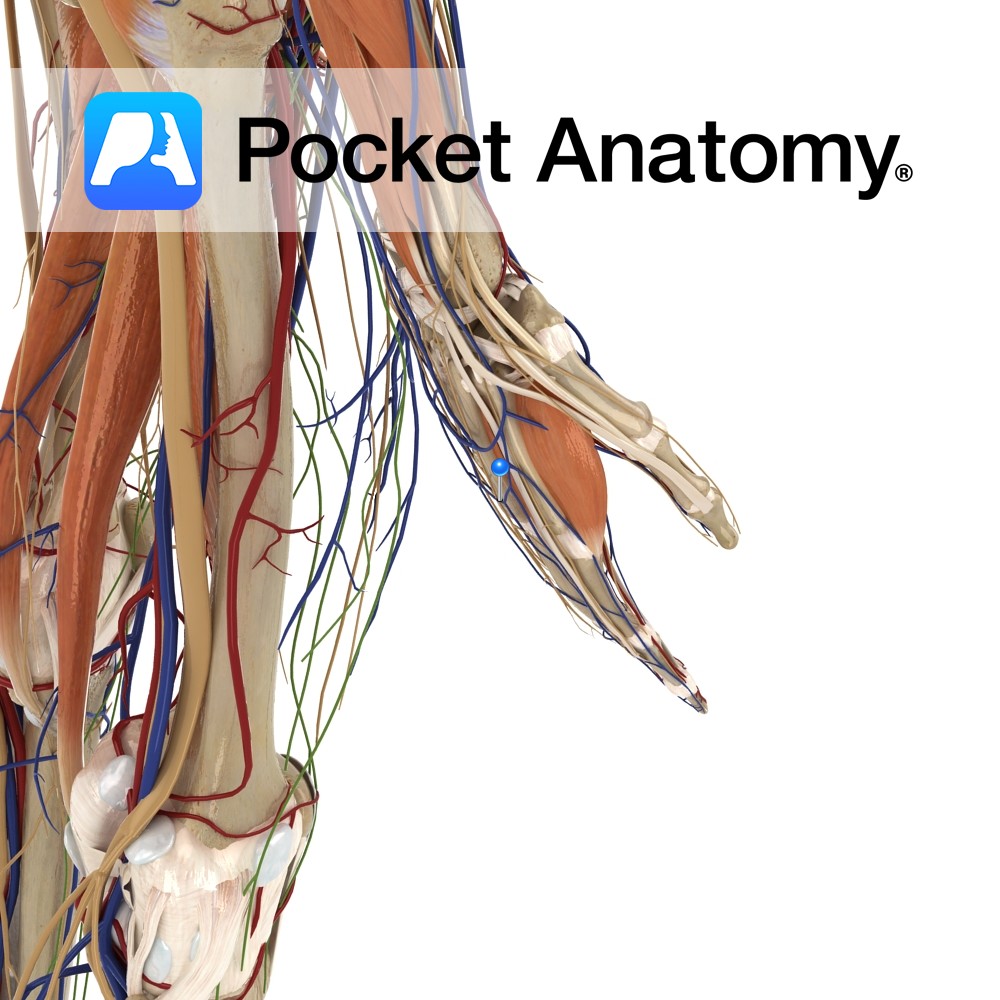
Anatomy Course A complex network of veins on the dorsum of the hand created by the dorsal metacarpal veins. The network gives rise to several veins, the most important being the cephalic and basilic vein. Drain Drains the dorsum of the hand. Interested in taking our award-winning Pocket Anatomy app for a test drive?
- Published in Pocket Anatomy Pins
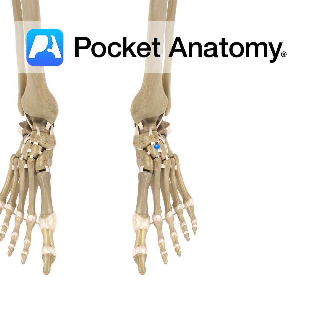
Anatomy Short, flat ligaments that attach longitudinally and obliquely from the cuboid and the three cuneiform bones, to the metatarsal bones of the foot. Functions Static stability to the tarsometatarsal joints. Clinical Injured when fractures or dislocations occur at the tarsometatarsal joint (at the first tarsometatarsal joint, called the lisfranc injury). Commonly injured when the
- Published in Pocket Anatomy Pins
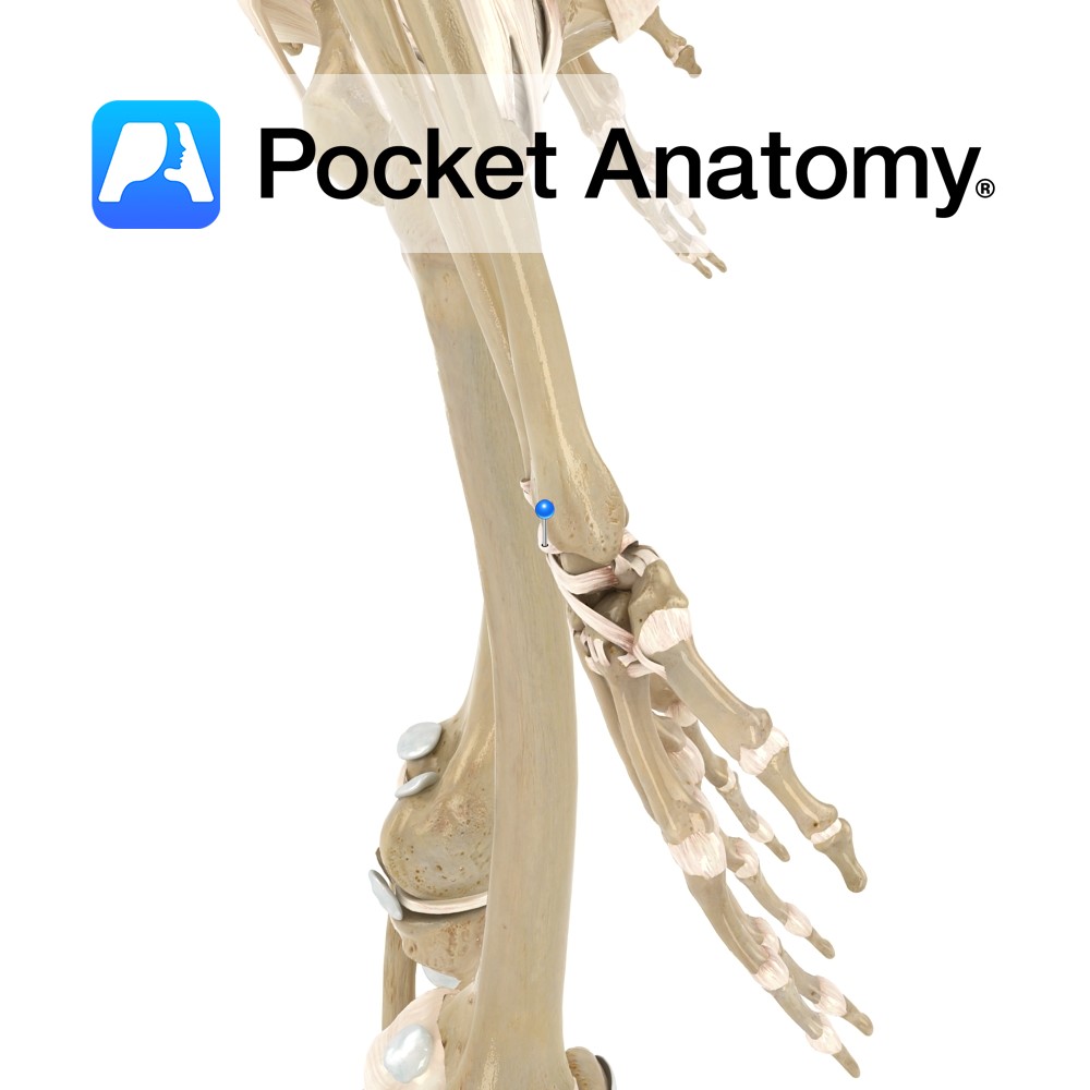
Anatomy Attaches to the posterior border of the distal radius. It travels obliquely downwards and medially to attach to the dorsal surface of the scaphoid, lunate and trapezium bones of the hand. Weaker than the volar radiocarpal ligament. Functions Helps to stabilize the wrist joint posteriorly. Interested in taking our award-winning Pocket Anatomy app for
- Published in Pocket Anatomy Pins
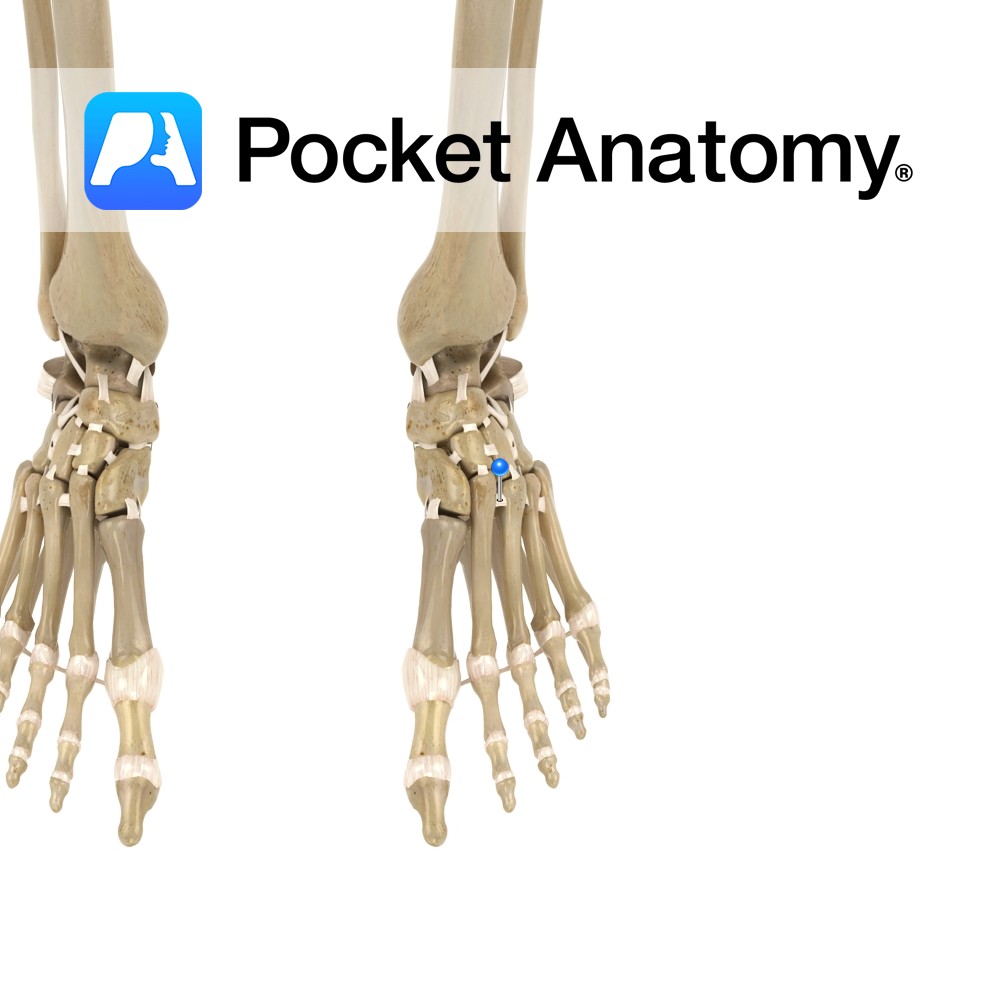
Anatomy A series of ligaments that attach from the dorsal surface of one metatarsal to the dorsal surface of the adjacent metatarsal. Situated at the proximal ends of the metatarsal bones. Functions Stabilizers of the metatarsal joints. Interested in taking our award-winning Pocket Anatomy app for a test drive?
- Published in Pocket Anatomy Pins
Anatomy Course First dorsal metatarsal artery is a direct branch of the dorsalis pedis artery. The second, third and fourth metatarsal arteries originate from the arcuate artery. Supply Together they supply the digits of the foot. Interested in taking our award-winning Pocket Anatomy app for a test drive?
- Published in Pocket Anatomy Pins
Anatomy Course Arise from the dorsal digital veins, which form three dorsal metacarpal veins. These travel briefly before creating a complex network on the dorsum of the hand. Drain Drain the dorsum of the hand. Interested in taking our award-winning Pocket Anatomy app for a test drive?
- Published in Pocket Anatomy Pins
Anatomy Thick bands of connective tissue attaching from the bases of the two to five metacarpal bones. Functions By linking metacarpal bones two to five, they help to form the skeletal framework of the palm. Interested in taking our award-winning Pocket Anatomy app for a test drive?
- Published in Pocket Anatomy Pins

