PocketAnatomy® is a registered brand name owned by © eMedia Interactive Ltd, 2009-2022.
iPhone, iPad, iPad Pro and Mac are trademarks of Apple Inc., registered in the U.S. and other countries. App Store is a service mark of Apple Inc.
Anatomy Origin: Plantar surface of the base of the 5th metatarsal and sheath of adjacent fibularis longus tendon. Insertion: Base of the 5th proximal phalanx. Key Relations: Runs along the lateral aspect of the foot. See plantar view of flexor digiti minimi brevis of foot.. Functions Flexes the 5th toe at metatarsophalangeal joint. Supply Nerve
- Published in Pocket Anatomy Pins
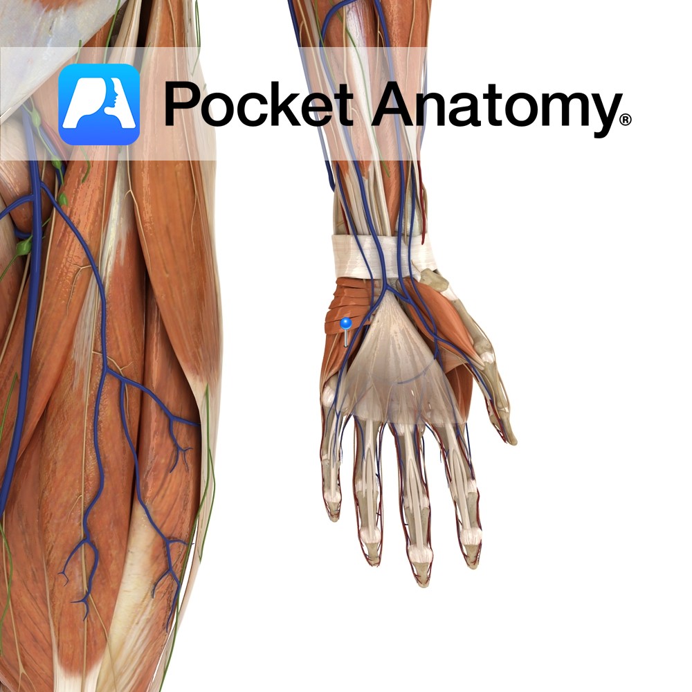
Anatomy Origin: Hook of the hamate and the ulnar border of the flexor retinaculum. Insertion: Ulnar aspect of the base of the proximal phalanx of the little finger. Key Relations: -Is one of the muscles of the hypothenar eminence of the hand. -Abductor digiti minimi lies on its lateral side and the two muscels are
- Published in Pocket Anatomy Pins
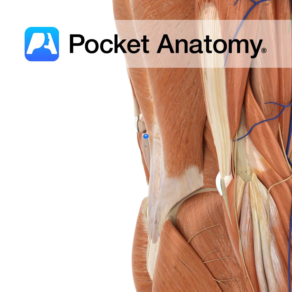
Anatomy Origin: Humeral head: Medial epicondyle of humerus via the common flexor tendon. Ulnar head: Medial margin of olecranon and upper two thirds of dorsal border of ulna by an aponeurosis. Insertion: Pisiform bone, then via pisohamate and pisometacarpal ligaments into the hook of the hamate and the fifth metacarpal. Key Relations: -The ulnar nerve
- Published in Pocket Anatomy Pins
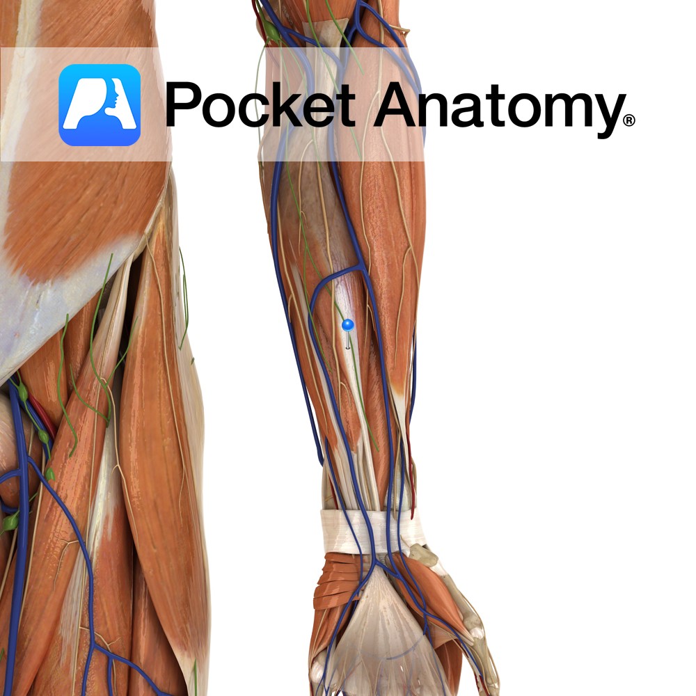
Anatomy Origin: Medial epicondyle of humerus via the common flexor tendon. Insertion: Plamar aspect of bases of second and third metacarpal bones (with a slip to the third). Key Relations: -The tendon of flexor carpi radialis is considered part of the carpal tunnel, although more accurately it passes through the flexor retinaculum, the cover of
- Published in Pocket Anatomy Pins
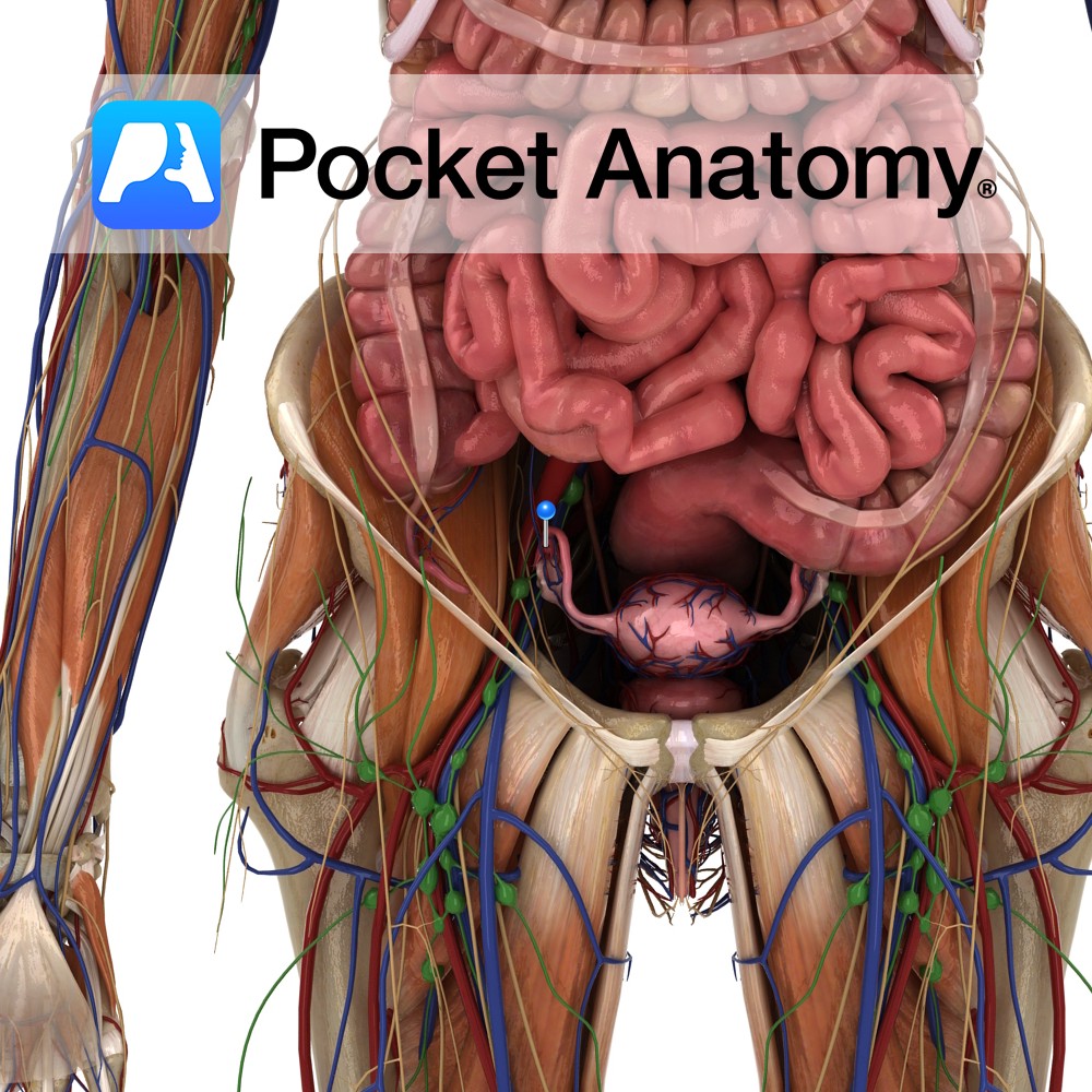
Anatomy Fringes/fingers projecting from widened lateral end (infundibulum) of fallopian tube, closely associated with ovary. One muscular fimbria – fimbria ovarica – is attached to the ovary. Fimbriae lined internally with millions of tiny hair-like cilia. Physiology Fimbria ovarica contracts at ovulation, pulling the tube more tightly towards the ovary. Cilia beat rapidly, creating current
- Published in Pocket Anatomy Pins
Anatomy Origin: Distal quarter of the anterior surface of the fibula and the interosseous membrane. Insertion: Dorsal surface of the base of the 5th metatarsal. Key relations: -One of the four muscles of the anterior compartment of the leg. -The fibularis tertius tendon passes posterior to the extensor retinaculae. It crosses anterior to the ankle
- Published in Pocket Anatomy Pins
Anatomy Origin: Head and upper two-thirds of the lateral surface of fibula and the intermuscular septa. Insertion: Lateral surface of the medial cuneiform and the base of the 1st metatarsal. Key relations: -One of the two muscles of the lateral compartment of the leg. -The fibularis longus tendon passes posterior to the lateral malleolus along
- Published in Pocket Anatomy Pins
Anatomy Origin: Distal two-thirds of the lateral surface of the fibula and the intermuscular septa. Insertion: Tuberosity on the lateral aspect of the base of the 5th metatarsal. Key Relations: -One of the two muscles of the lateral compartment of the leg. -The fibularis brevis tendon passes posterior to the lateral malleolus along the lateral
- Published in Pocket Anatomy Pins
Anatomy Course Branch of the posterior tibial artery, which follows a parallel course to the tibial artery on the lateral side of the posterior fibula. Supply Supplies muscles and bones in the posterior compartment of the leg, but also the lateral compartment via its branches. Interested in taking our award-winning Pocket Anatomy app for a
- Published in Pocket Anatomy Pins
Anatomy The shaft is connected to that of tibia along its length by an interossous membrane, separating anterior and posterior muscles. The membrane is 1 of 3 articulations between tibia and fibula, along with the proximal tibiofibular joint above and the tibiofibular syndesmosis below. Clinical Fracture of tibia usually associated with fracture fibula, as interosseous
- Published in Pocket Anatomy Pins

.jpg)
-tertius.jpg)
-longus.jpg)
-brevis.jpg)
-artery.jpg)
