PocketAnatomy® is a registered brand name owned by © eMedia Interactive Ltd, 2009-2022.
iPhone, iPad, iPad Pro and Mac are trademarks of Apple Inc., registered in the U.S. and other countries. App Store is a service mark of Apple Inc.
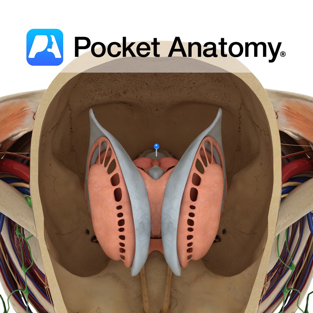
Anatomy Is a rhomboid or diamond shaped cavity lined with ependymal cells part of the series of fluid-filled cavities, which make up the ventricular system. It is located anterior to the cerebellum and posterior to the pons and superior half of the medulla. It is connected superiorly to the third ventricle via the cerebral aqueduct
- Published in Pocket Anatomy Pins
Motion There are two radioulnar joints: the proximal radioulnar joint and distalradioulnar joint. Both are synovial pivot joints. They permit pronation and supination only. Proximal radioulnar joint: The circumference of the radial head articulates with the fibro-osseous ring made by the ulnar radial notch and annular ligament. Distal radioulnar joint: The distal head of the
- Published in Pocket Anatomy Pins
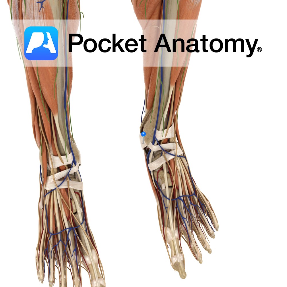
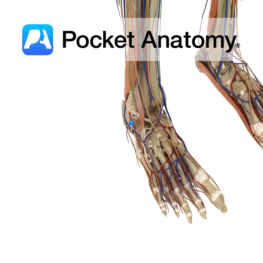
Anatomy Attaches from the posterior surface of the medial malleolus, pacing obliquely backwards to the inferiomedial margin of the calcaneus. Functions Provides static stability to the medial ankle joint. Acts as a pulley for the flexor halluces longus, flexor digitorum longus and tibials posterior tendons. Vessels of the foot pass under the flexor retinaculum. Clinical
- Published in Pocket Anatomy Pins
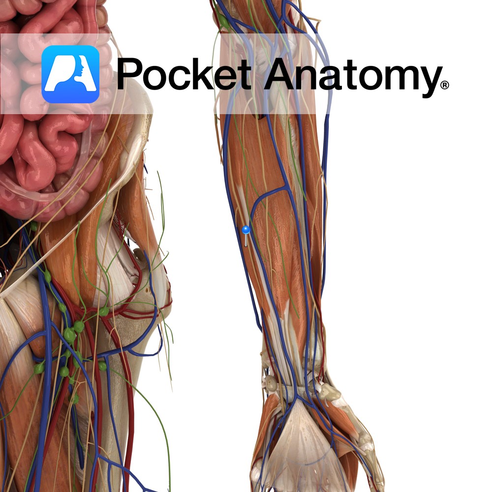
Anatomy Origin: Anterior surface of middle third of radius and adjacent interosseous membrane. Insertion: Base of the distal phalanx of thumb. Key Relations: -The tendons of flexor pollicis longus passes through the carpal tunnel as it enter the palm of the hand. -One of the three muscles in the deep anterior compartment of the forearm.
- Published in Pocket Anatomy Pins
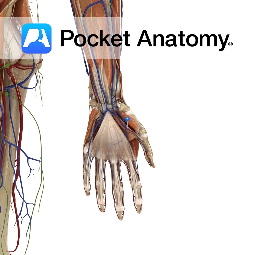
Anatomy Origin: Superficial part: Flexor retinaculum and tubercle of the trapezium. Deep part: Trapezoid and capitates. Insertion: Base of the proximal phalanx of thumb. Key Relations: Is one of the muscles of the thenar eminence of the hand. Functions Flexion of the thumb at the metacarpophalangeal joint. Supply Nerve Supply: Superficial part: Recurrent branch of
- Published in Pocket Anatomy Pins
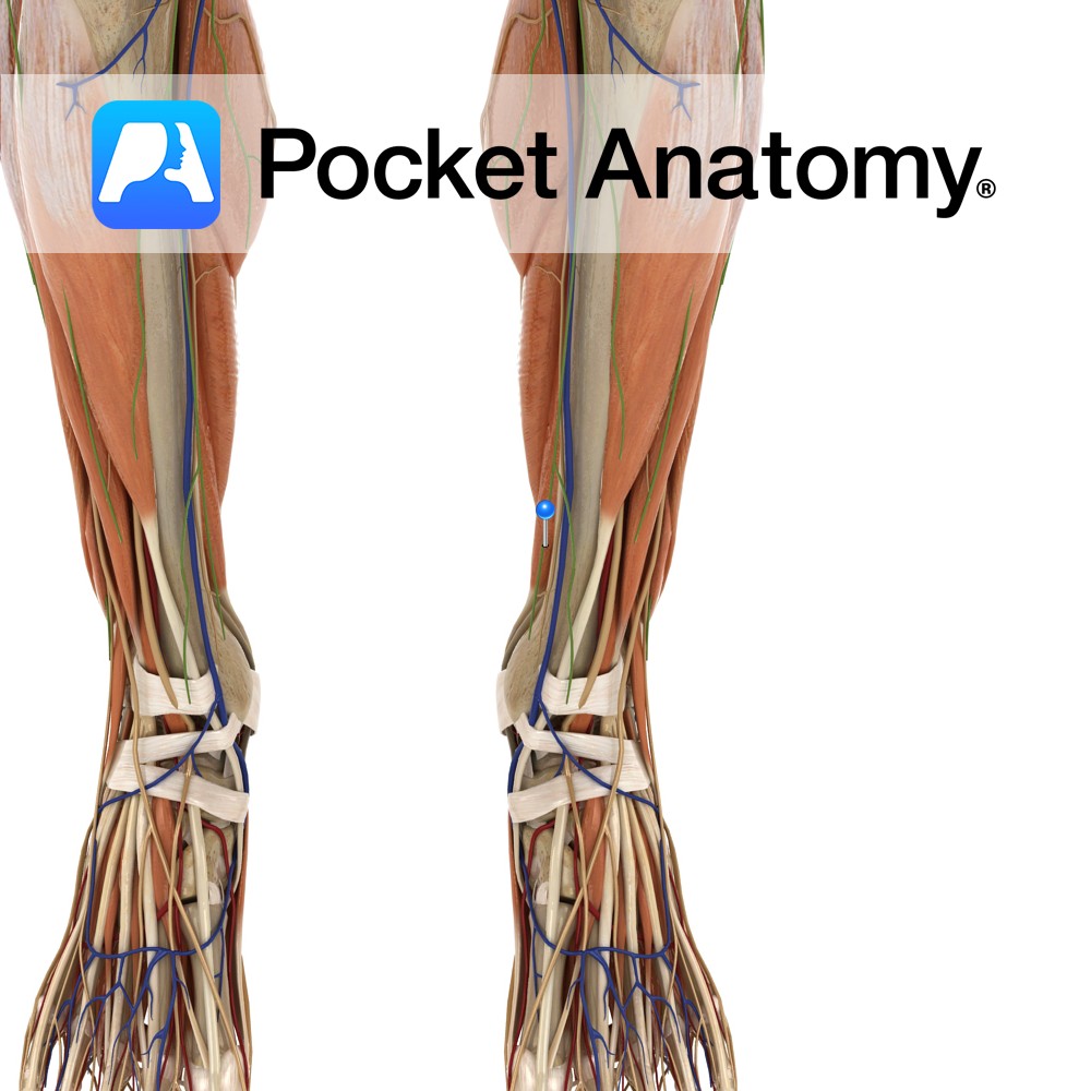
Anatomy Origin: Lower two-thirds of the posterior surface of the fibula, the posterior intermuscular septum and the posterior surface of the distal part of the interosseous membrane. Insertion: Plantar surface of the base of the distal phalanx of the hallux (big toe). Key relations: -One of the four muscles of the deep posterior compartment of
- Published in Pocket Anatomy Pins
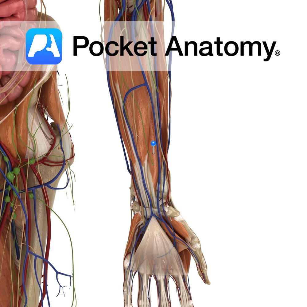
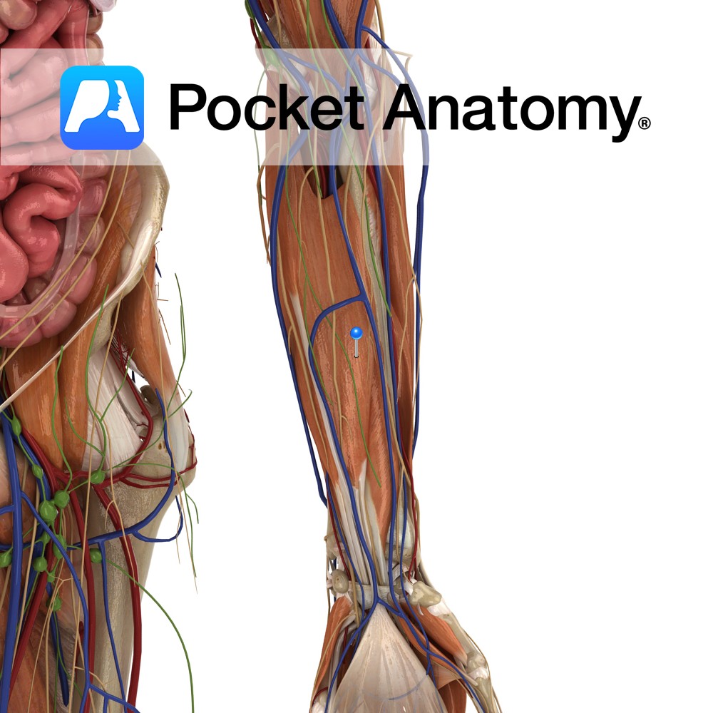
Anatomy Origin: Humeroulnar head: Medial epicondyle of humerus via the common flexor tendon, ulnar collateral ligament and coronoid process of ulna. Radial head: Superior half of anterior aspect of radius at anterior oblique line. Insertion: Four tendons of insertion attach to palmar surfaces of bodies of middle phalanges of the four fingers. Key Relations: -The
- Published in Pocket Anatomy Pins
Anatomy Origin: Upper three quarters of medial and anterior aspects of ulna and from anterior aspect of interosseous membrane. Insertion: Four tendons attaching to palmar aspects of bases of the distal phalanges of each of the four fingers. Key Relations: -The lumbrical muscles arise in the palm from the tendons of flexor digitorum profundus. -The
- Published in Pocket Anatomy Pins
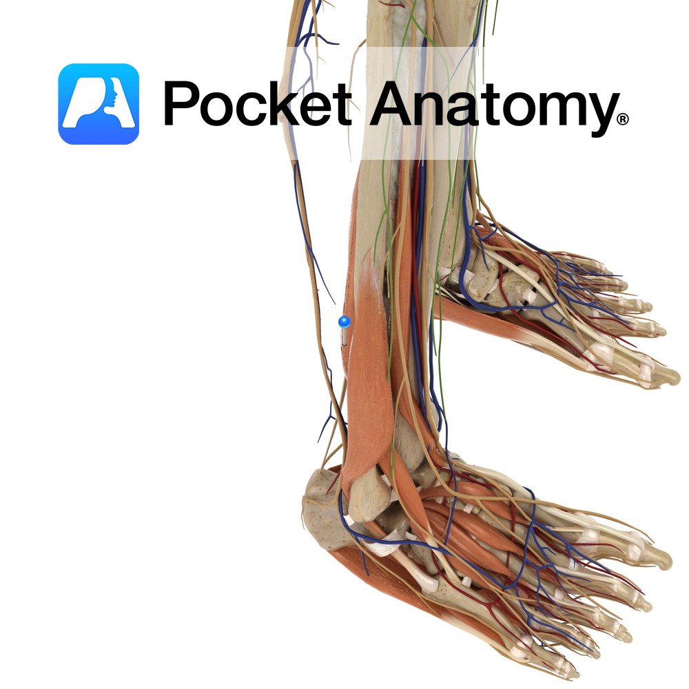
Anatomy Origin: Medial side of the posterior surface of the tibia inferior to the soleal line. Insertion: Plantar surface of the base of the distal phalanges of the lateral four toes. Key Relations: -One of the four muscles of the deep posterior compartment of the leg. -The flexor digitorum longus tendon passes posterior to the
- Published in Pocket Anatomy Pins
Anatomy Origin: Medial process of the calcaneal tuberosity and plantar aponeurosis. Insertion: Sides of the plantar surface of the middle phalanges of the lateral 4 toes. Key relations: -Lies superior to the plantar aponeurosis. -Gives rise to 4 tendons that lie superficial to the flexor digitorum longus tendons. See plantar view of flexor digitorum brevis.
- Published in Pocket Anatomy Pins

.jpg)
