PocketAnatomy® is a registered brand name owned by © eMedia Interactive Ltd, 2009-2022.
iPhone, iPad, iPad Pro and Mac are trademarks of Apple Inc., registered in the U.S. and other countries. App Store is a service mark of Apple Inc.
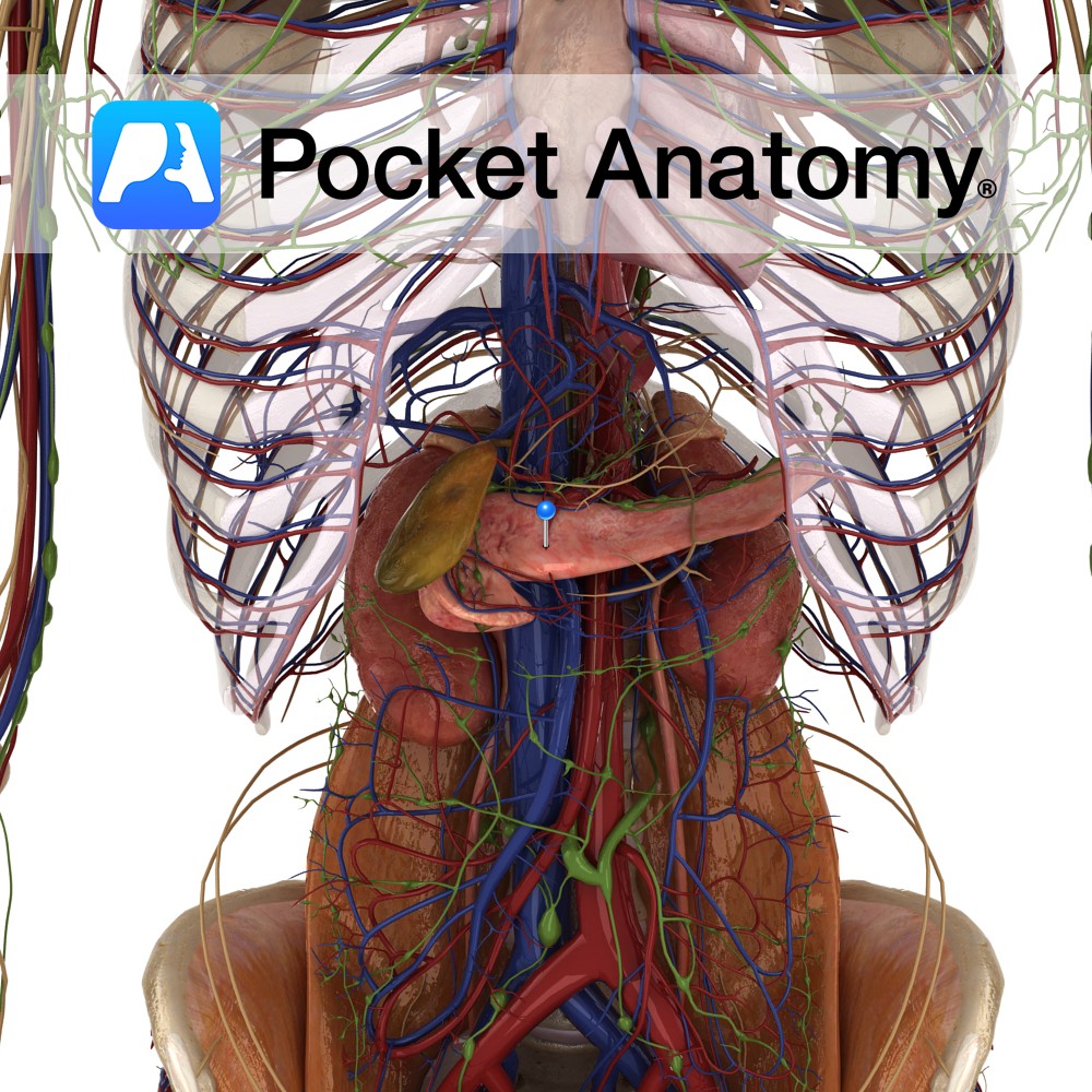
Anatomy Course Branch of the hepatic artery that descends posterior to the duodenum until it reaches its inferior border and gives off its terminal branches. Supply Supplies the pylorus of the stomach and the proximal duodenum. Interested in taking our award-winning Pocket Anatomy app for a test drive?
- Published in Pocket Anatomy Pins
Anatomy Origin: Medial head: Popliteal surface above the medial condyle of the femur. Lateral head: Lateral aspect of the lateral condyle of the femur. Insertion: Middle part of the posterior surface of the calcaneus by the tendo calcaneus (Achilles tendon). Key Relations: -One of the three muscles of the superficial posterior compartment of the leg.
- Published in Pocket Anatomy Pins
Anatomy Origin: Lateral head: Lateral aspect of the lateral condyle of the femur. Medial head: Popliteal surface above the medial condyle of the femur. Insertion: Middle part of the posterior surface of the calcaneus by the tendo calcaneus (Achilles tendon). Key Relations: -One of the three muscles of the superficial posterior compartment of the leg.
- Published in Pocket Anatomy Pins
Anatomy Hollow, muscular, very distensible, pear-shaped organ under liver; receives bile through common hepatic, cystic ducts; concentrates (strengthens) and reserves; releases into cystic duct, on to common bile duct, ampulla of Vater, duodenum through papilla; c. 3″ long, 4″ diameter full; fundus, body, neck (sometimes with saccular Hartman’s pouch), tapering to duct; inferior covered by
- Published in Pocket Anatomy Pins
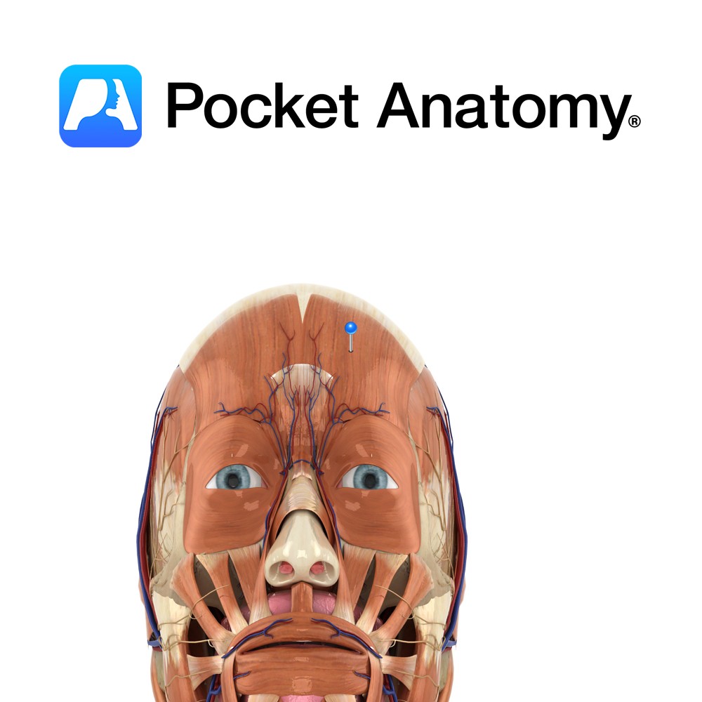
Anatomy Origin: Frontalis has no bony origin. Its fibres are continuous with those of the muscles procerus and corrgator supercilii. Insertion: Fibres join the galea aponeurotica (epicranial aponeurosis) below the coronal suture. Key Relations: Many consider frontalis and occipitalis as one muscle (called occipitofrontalis) with an aponeurotic tendon between two bellies- a frontal belly and
- Published in Pocket Anatomy Pins
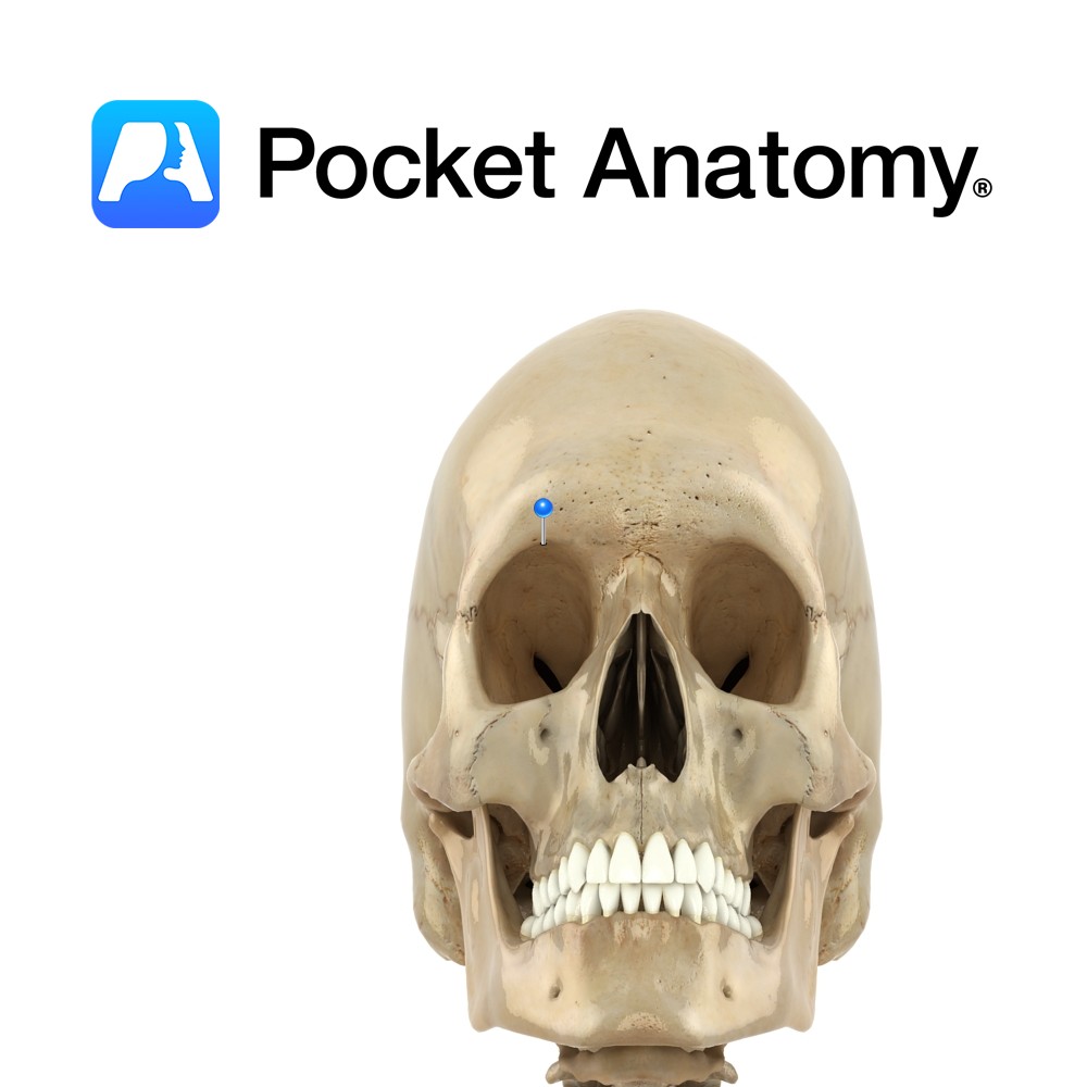
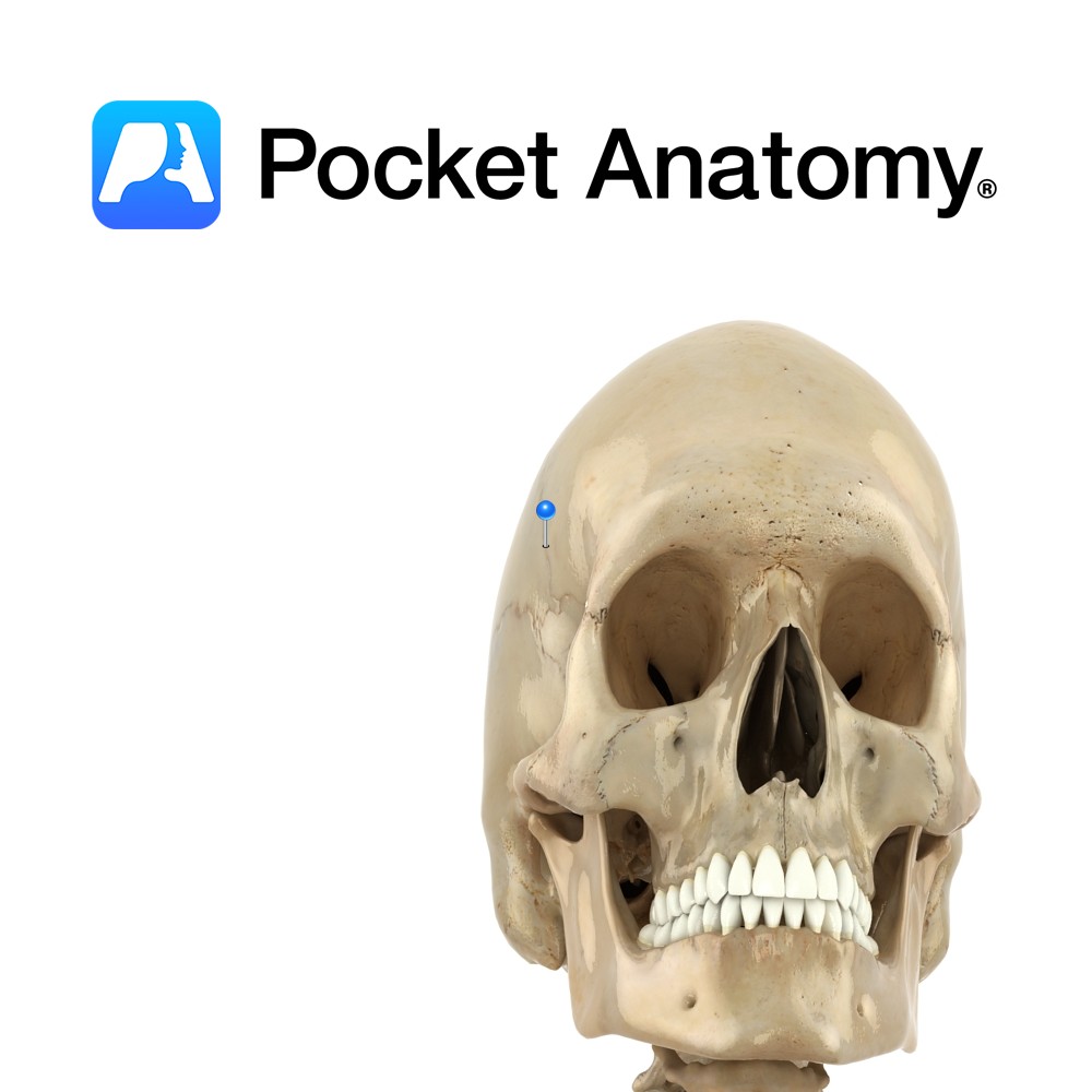
Anatomy Small notch at medial margin on top of each eye socket, through which supra-orbital vessels and nerve pass through. Can be a foramen or a notch. Vignette Swimmer’s headache may be caused by tight goggles pressing on supraorbital nerve at (or near) notch. Interested in taking our award-winning Pocket Anatomy app for a test
- Published in Pocket Anatomy Pins
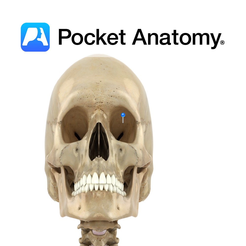
Anatomy Section of frontal bone which forms part of eye sockets/orbits. Frontal bone curves in at bottom, as thin triangular orbital plates. Left and right plate are separated by a gap – the ethmoid notch. Interested in taking our award-winning Pocket Anatomy app for a test drive?
- Published in Pocket Anatomy Pins
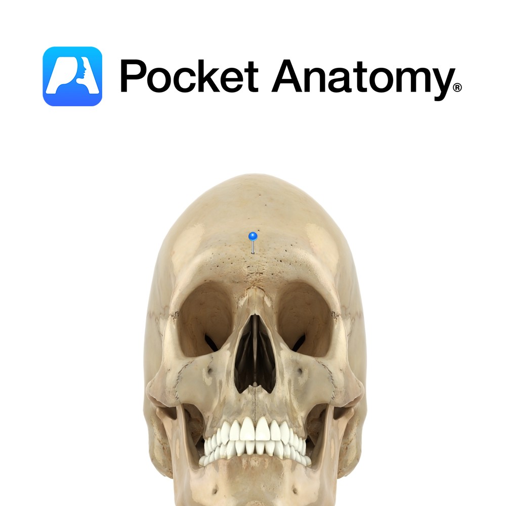
Anatomy Slightly raised flat area where the superciliary arches meet (ie between eyebrows); the most forward part of the forehead. Clinical Glabellus (Latin); smooth. Usually hairless. Vignette Site for testing the Glabellar reflex; tapping on glabella causes blinking, which stops after several repetitions. However, often persists in Parkinson’s (Myerson’s sign). Interested in taking our award-winning
- Published in Pocket Anatomy Pins
Anatomy Rigid midline joint or seam between frontal bone and paired parietal bones behind. Ends at side of skull at junction of frontal, parietal and sphenoid bones. Vignette Korone (Greek); garland, wreath. Interested in taking our award-winning Pocket Anatomy app for a test drive?
- Published in Pocket Anatomy Pins
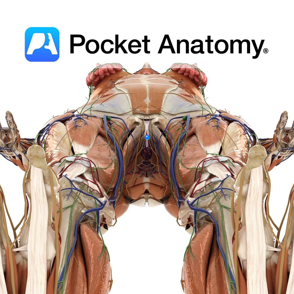
Anatomy Otherwise referred to as the fourchette, this is the meeting of the labia minora behind the vagina, in front of the anus (their anterior meeting/junction swaddles the clitoris as prepuce and frenulum). It is part of the vulva (external genital organs – including mons, clitoris, urethra/vaginal orifices, labia majora/minora), itself part of the perineum
- Published in Pocket Anatomy Pins

.jpg)
.jpg)

