PocketAnatomy® is a registered brand name owned by © eMedia Interactive Ltd, 2009-2022.
iPhone, iPad, iPad Pro and Mac are trademarks of Apple Inc., registered in the U.S. and other countries. App Store is a service mark of Apple Inc.
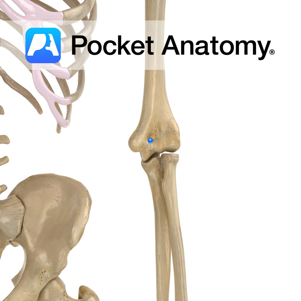
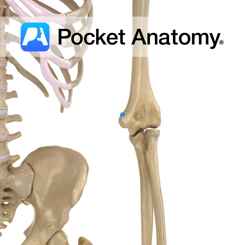
Anatomy The articular surface of the bottom of humerus is divided into trochlea medially (for trochlear notch of ulna, which is between coronoid process and olecranon) and capitulum laterally (for head of radius). Vignette Trochlea (Latin); pulley. Interested in taking our award-winning Pocket Anatomy app for a test drive?
- Published in Pocket Anatomy Pins

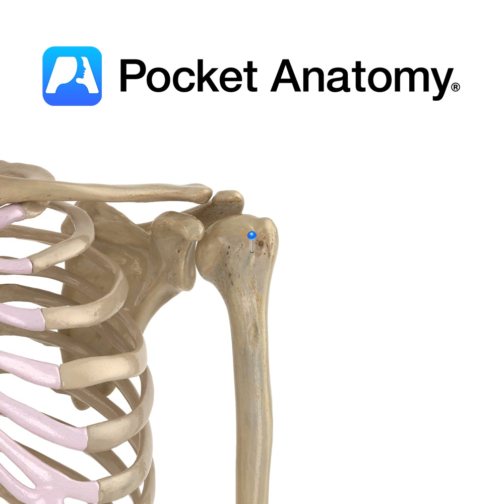
Anatomy Common fracture site (much more so than anatomical neck), with risk of damage to adjacent axillary nerve. Interested in taking our award-winning Pocket Anatomy app for a test drive?
- Published in Pocket Anatomy Pins
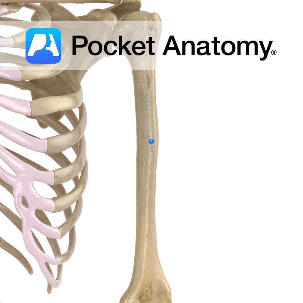
Anatomy Upper part cylindrical, lower prism-shaped. Deltoid tuberosity (for attachment of deltoid muscle) on antero-lateral aspect (to outside and front) at mid portion shaft. Interested in taking our award-winning Pocket Anatomy app for a test drive?
- Published in Pocket Anatomy Pins
Anatomy Just above the capitulum (which articulates with head of radius) at bottom of humerus laterally. When the arm is bent (ie elbow hinge joint flexed), the front part of radial head sits in it (limiting further flexion). Interested in taking our award-winning Pocket Anatomy app for a test drive?
- Published in Pocket Anatomy Pins
Anatomy When arm straightened (ie elbow hinge joint fully extended), the olecranon process of ulna sits in olecranon fossa at back of bottom of humerus, lending significant bony strength, stability and movement limitation (stopping hyperextension). Interested in taking our award-winning Pocket Anatomy app for a test drive?
- Published in Pocket Anatomy Pins
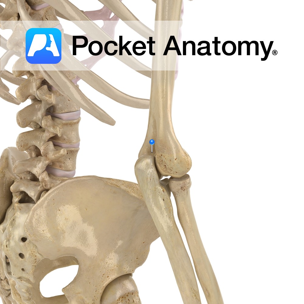
Anatomy Bump at bottom of humerus, medially (when hand pronated, ie palm forward). Attached; ulnar collateral ligament of elbow (significant in stabilizing the joint against valgus – lateral flexion), pronator teres, common flexor tendon. Bigger than lateral epicondyle. Clinical “Funny bone”; ulnar nerve runs just behind it (and can be easily felt – cordlike), with
- Published in Pocket Anatomy Pins
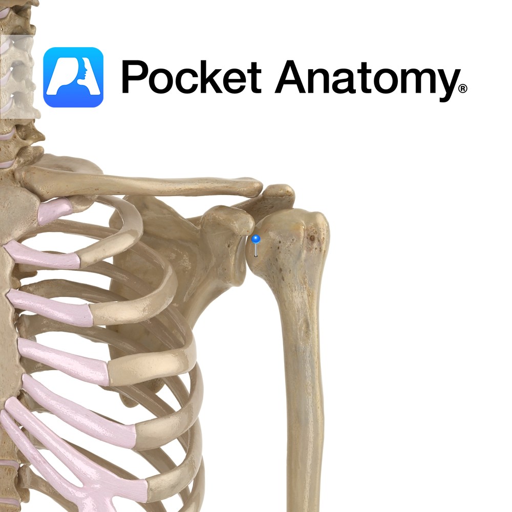
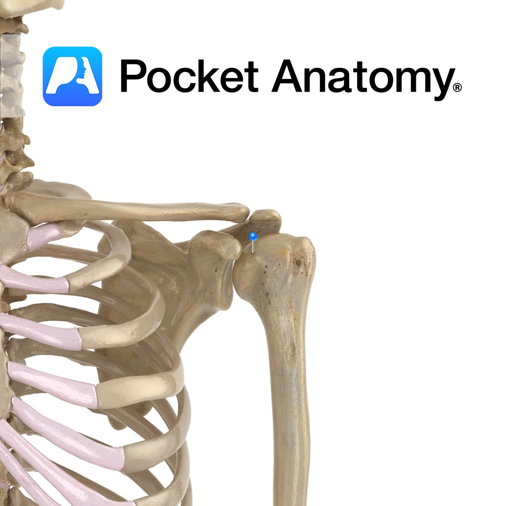
Anatomy Swelling below and medial to humeral head, below and in front of greater tubercle. Attachment; subscapularis. Interested in taking our award-winning Pocket Anatomy app for a test drive?
- Published in Pocket Anatomy Pins
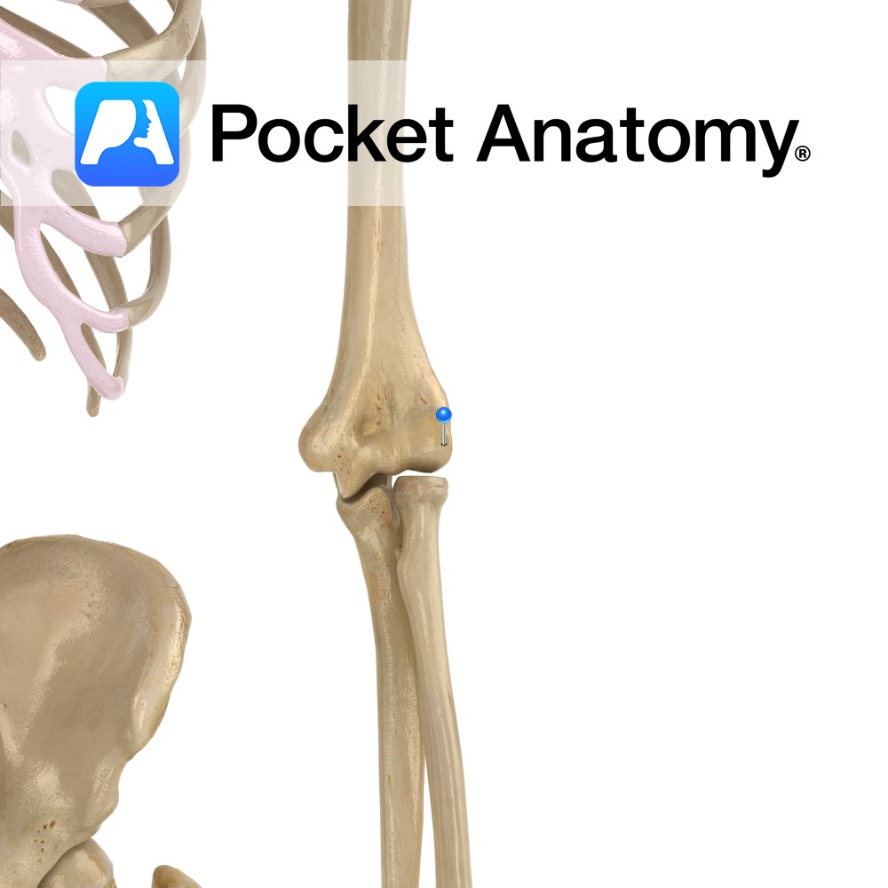
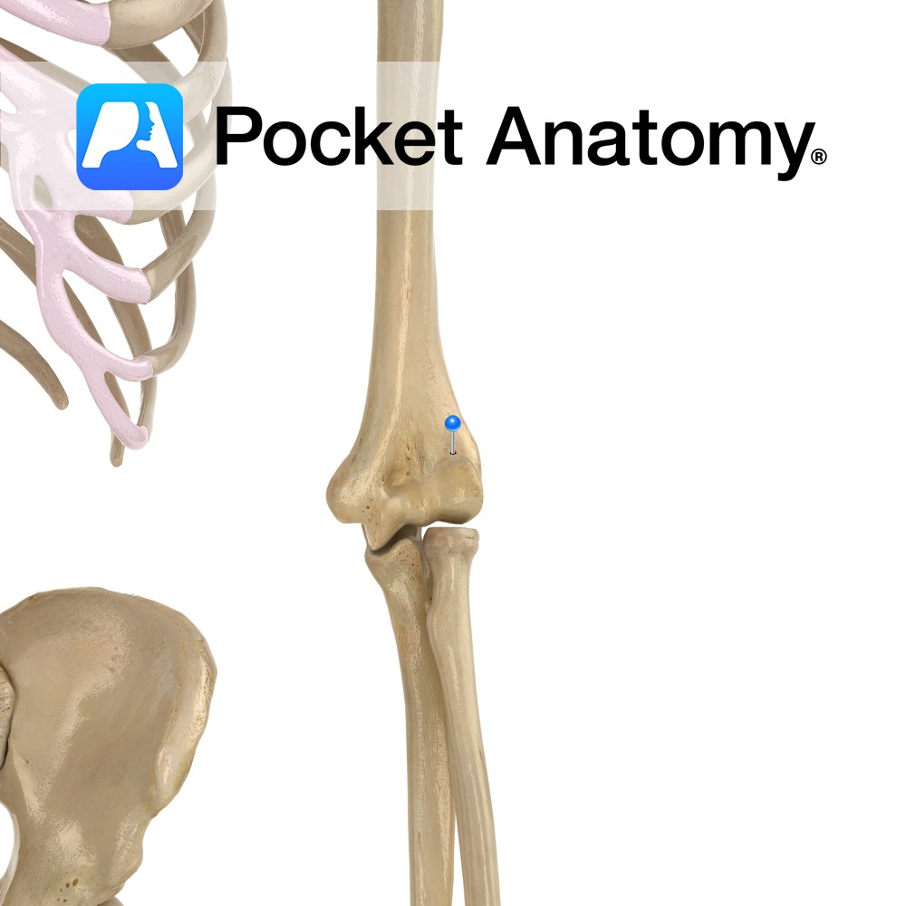
Anatomy Bump at bottom of humerus, laterally (when hand pronated, ie palm forward). Attached; radial collateral ligament (contributes to stabilizing the joint against varus – medial flexion), supinator, extensors. Smaller than medial epicondyle. Clinical Site of tennis elbow. Supercondylar fractures common in children; anterior displacement of upper part of fracture (proximal fragment) can damage brachial
- Published in Pocket Anatomy Pins
Anatomy Groove between greater and lesser tubercles, running down upper 1/3 of shaft of humerus. Attachment; long tendon latissimus dorsi (teres major attached medially, pectoralis major laterally). Interested in taking our award-winning Pocket Anatomy app for a test drive?
- Published in Pocket Anatomy Pins
Anatomy Rounded upper extremity of humerus; articulates with scapula at glenoid cavity/fossa. Vignette Shoulder is most mobile joint; flexion 150-170°, extension 40°, abduction 160-180°, adduction 30-40°, lateral rotation (in abduction) 95° (in adduction) 70°; medial rotation (in abduction) 40-50° (in adduction) 70°. Interested in taking our award-winning Pocket Anatomy app for a test drive?
- Published in Pocket Anatomy Pins