PocketAnatomy® is a registered brand name owned by © eMedia Interactive Ltd, 2009-2022.
iPhone, iPad, iPad Pro and Mac are trademarks of Apple Inc., registered in the U.S. and other countries. App Store is a service mark of Apple Inc.
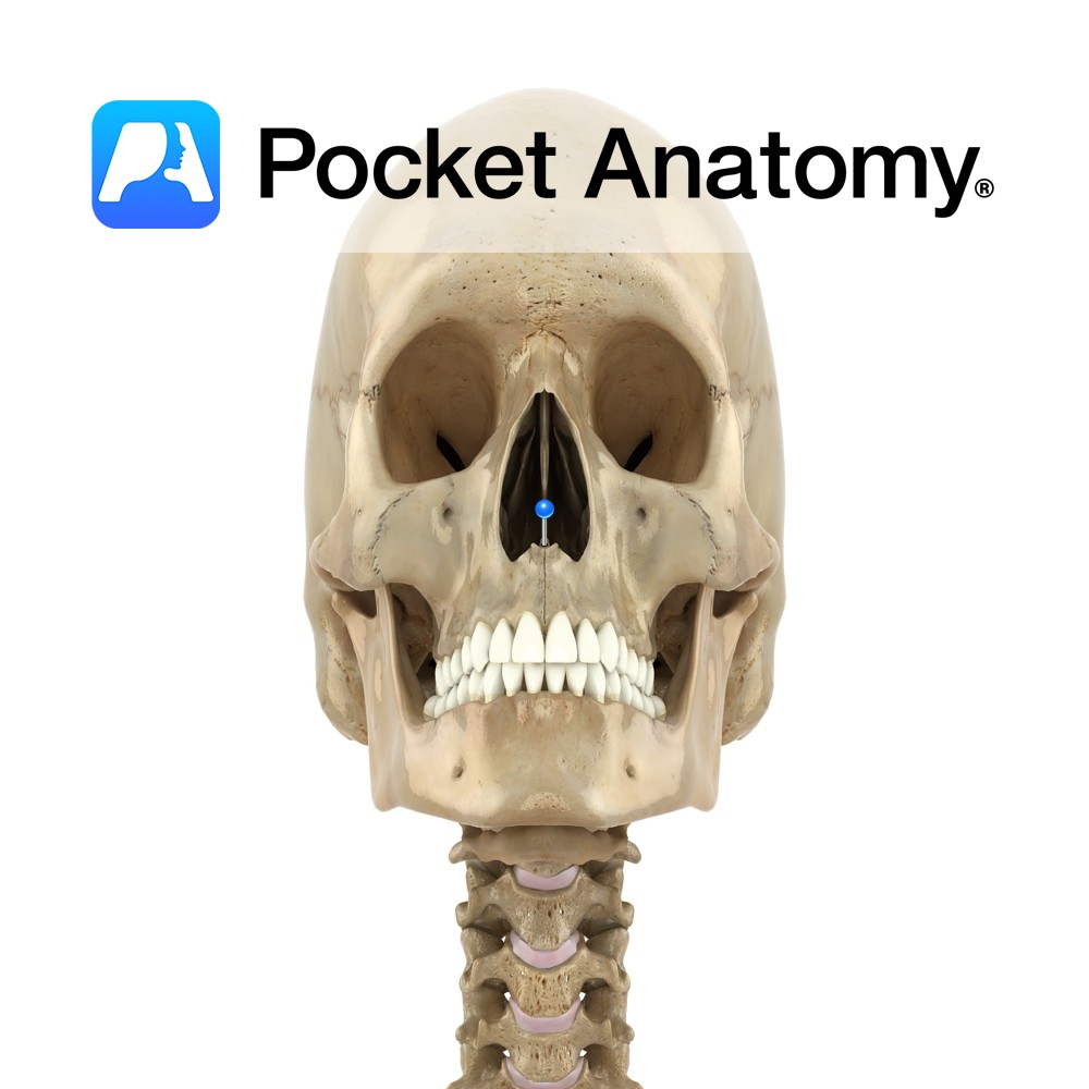
Anatomy The pointed lower ends (left and right) of the medial concave nasal notches of the frontal processes of the maxillae (4 processes; zygomatic, frontal, alveolar, palatine), meet and form this spine. Supports the lower back part of the septal cartilage. Vignette Can be palpated (felt) at junction of bottom of nose and philtrum (midline
- Published in Pocket Anatomy Pins
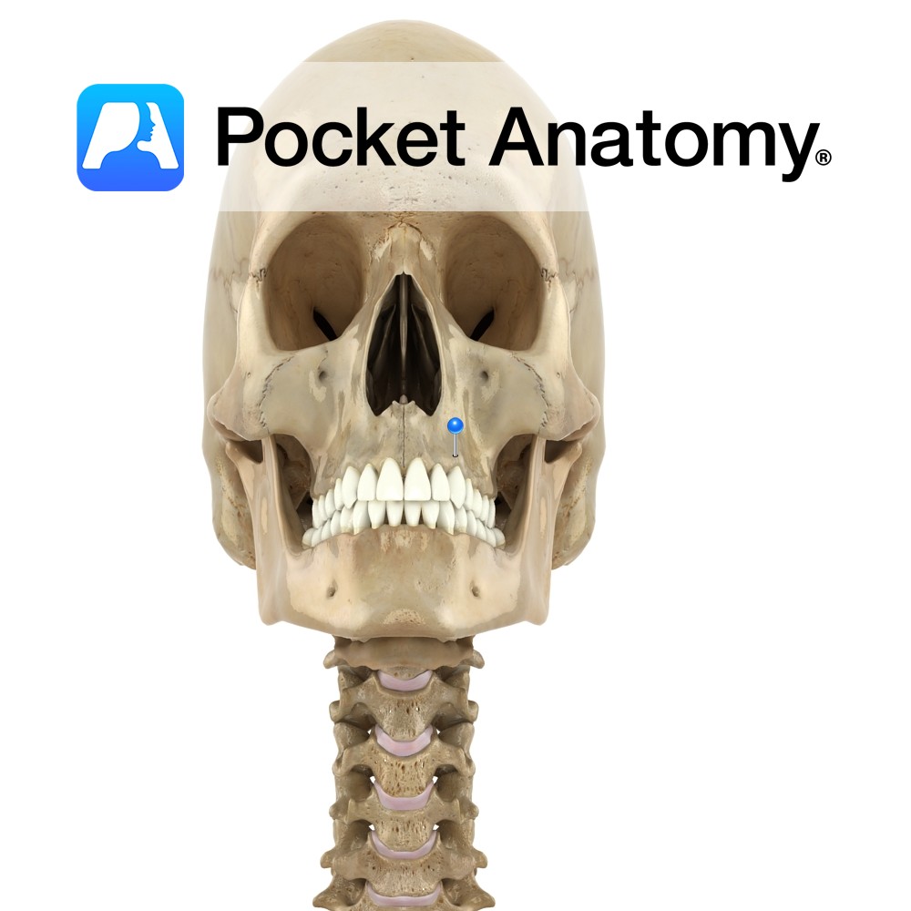
Anatomy Thick ridge of spongy bone, with cavities (tooth sockets) for 16 upper teeth (8×2). Interested in taking our award-winning Pocket Anatomy app for a test drive?
- Published in Pocket Anatomy Pins
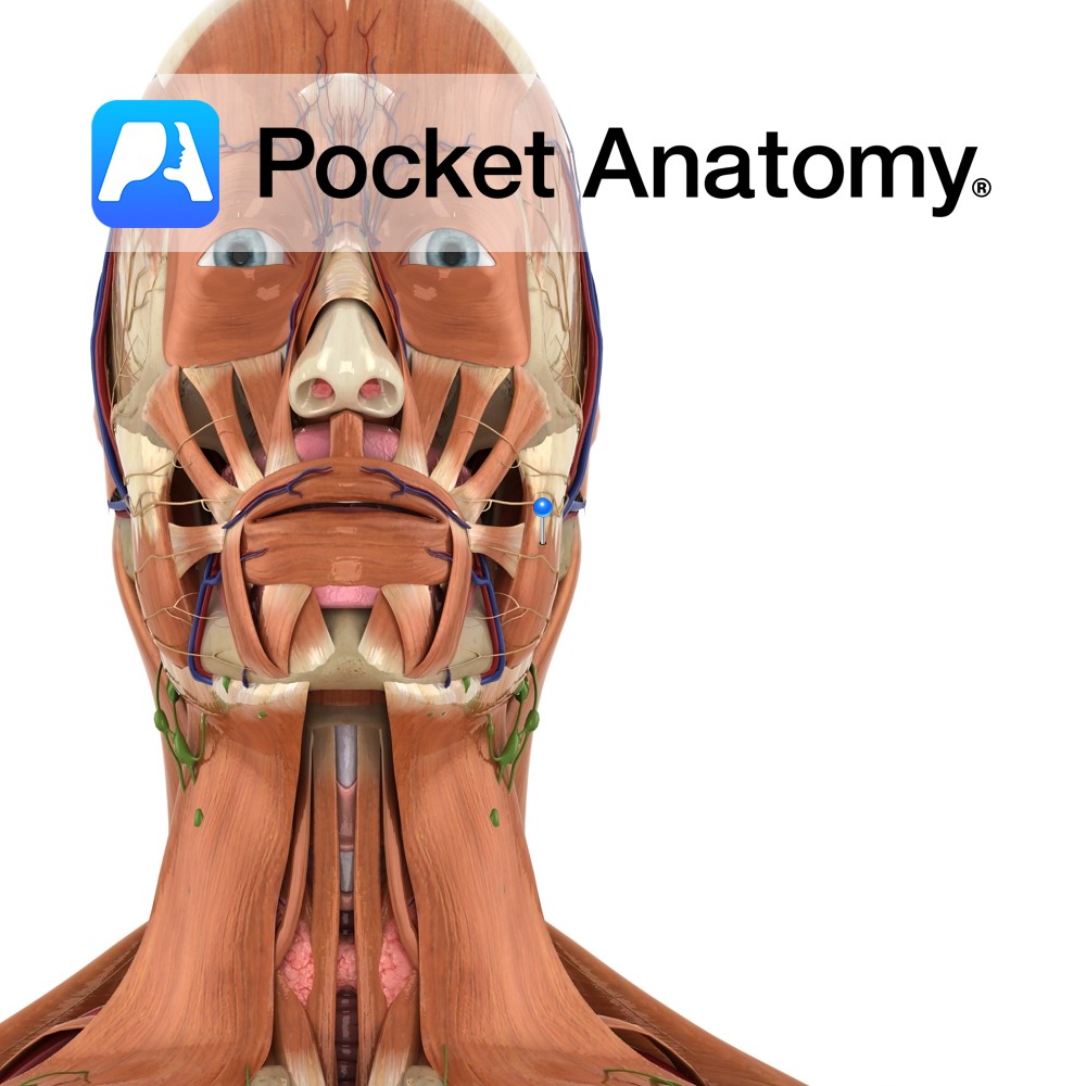
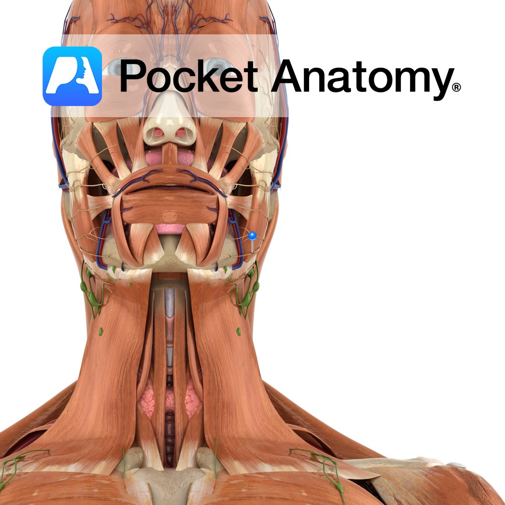
Anatomy Origin: Superficial and Deep portion: Inferior border and medial surface of zygomatic arch. Insertion: Superficial and Deep portion: Outer aspect of angle of jaw and lower half of ramus of mandible. Key Relations: -The superficial portion is larger and thicker whereas the deep portion is much smaller. -The muscle is crossed superficially by the
- Published in Pocket Anatomy Pins
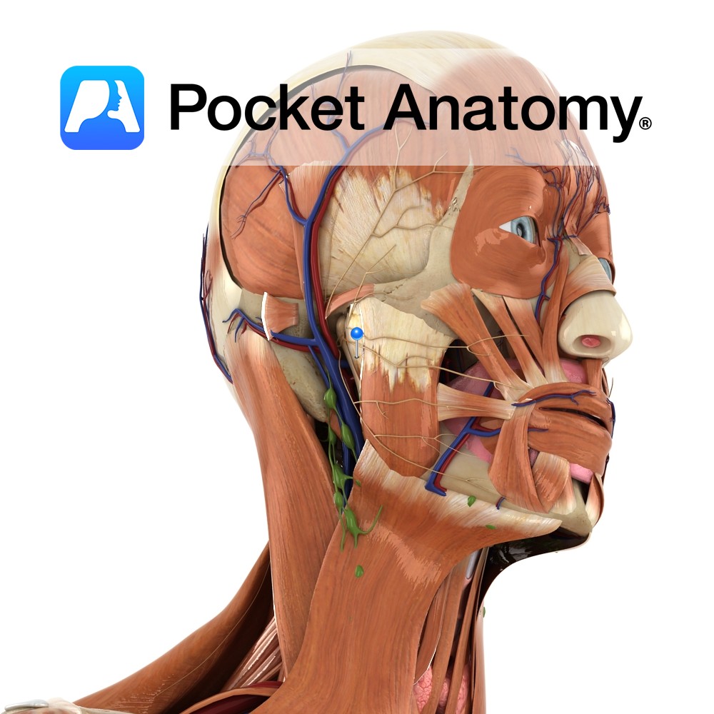
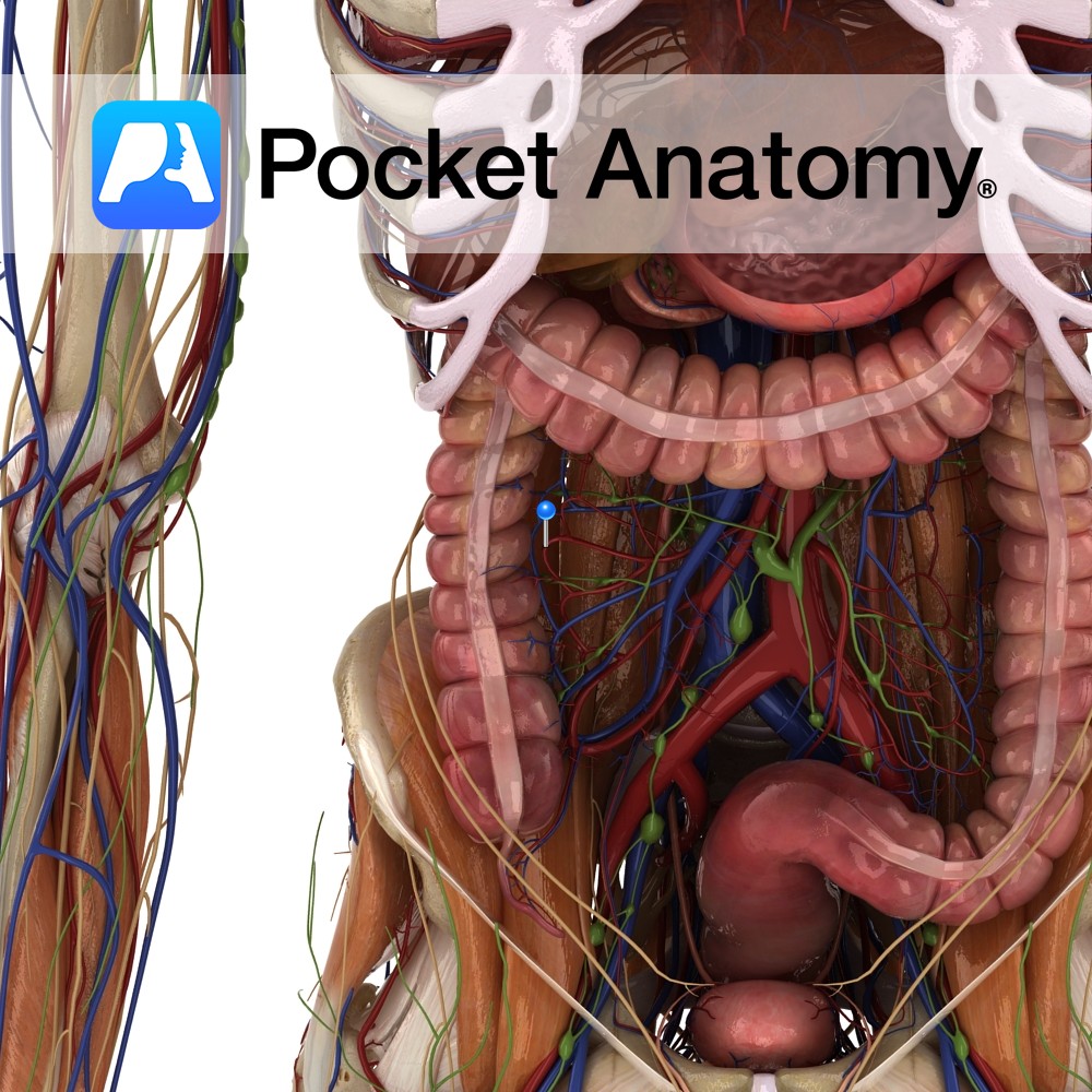
Anatomy Course Connects the superior and inferior mesenteric arteries in the gut. Formed when an anastomosis forms between the right, middle and left colic arteries, as well as the ileocolic artery. It runs in the mesentery with the other vessels of the bowel. Supply Supplies the bowel with blood, in conjunction with the other branches
- Published in Pocket Anatomy Pins
Anatomy Branch of the facial nerve (also known as the seventh cranial nerve). Facial nerve: Has a motor and sensory origin that join together to form the nerve. It passes through the internal auditory meatus through the facial canal and finally exits from the stylomastoid foramen, and into the parotid gland where it divides into
- Published in Pocket Anatomy Pins
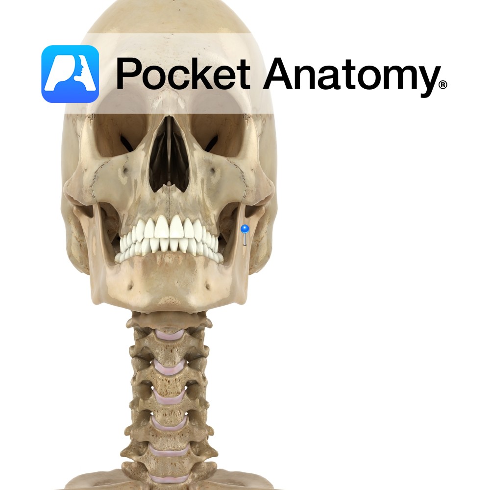
Anatomy The perpendicular part of the mandible, its upper extremities consisting of the coronoid and condylar processes, with the mandibular notch between. Laterally, almost fully covered by masseter; postero-medially, pterygoid. Clinical The mandible (jawbone) holds the lower teeth. Chews. involved in speech. Along with cranium, makes up skull. Four parts (each side): Anterior (left and
- Published in Pocket Anatomy Pins
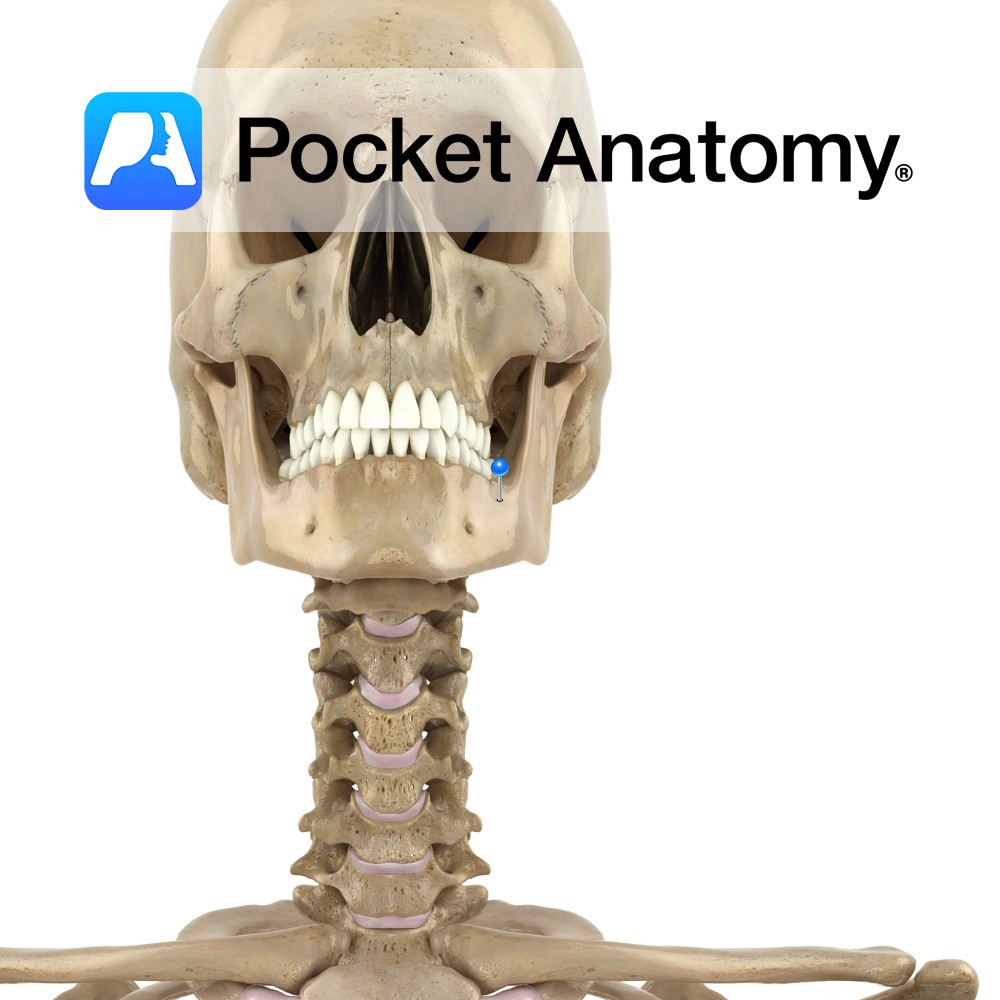
Anatomy Consists of a ridge. Begins faintly anteriorly where it begins at mental tubercle (chin), more evident as it courses back and up to become continuous with anterior border ramus. Clinical Attachment for depressor anguli oris, quadratus labii inferioris. Interested in taking our award-winning Pocket Anatomy app for a test drive?
- Published in Pocket Anatomy Pins
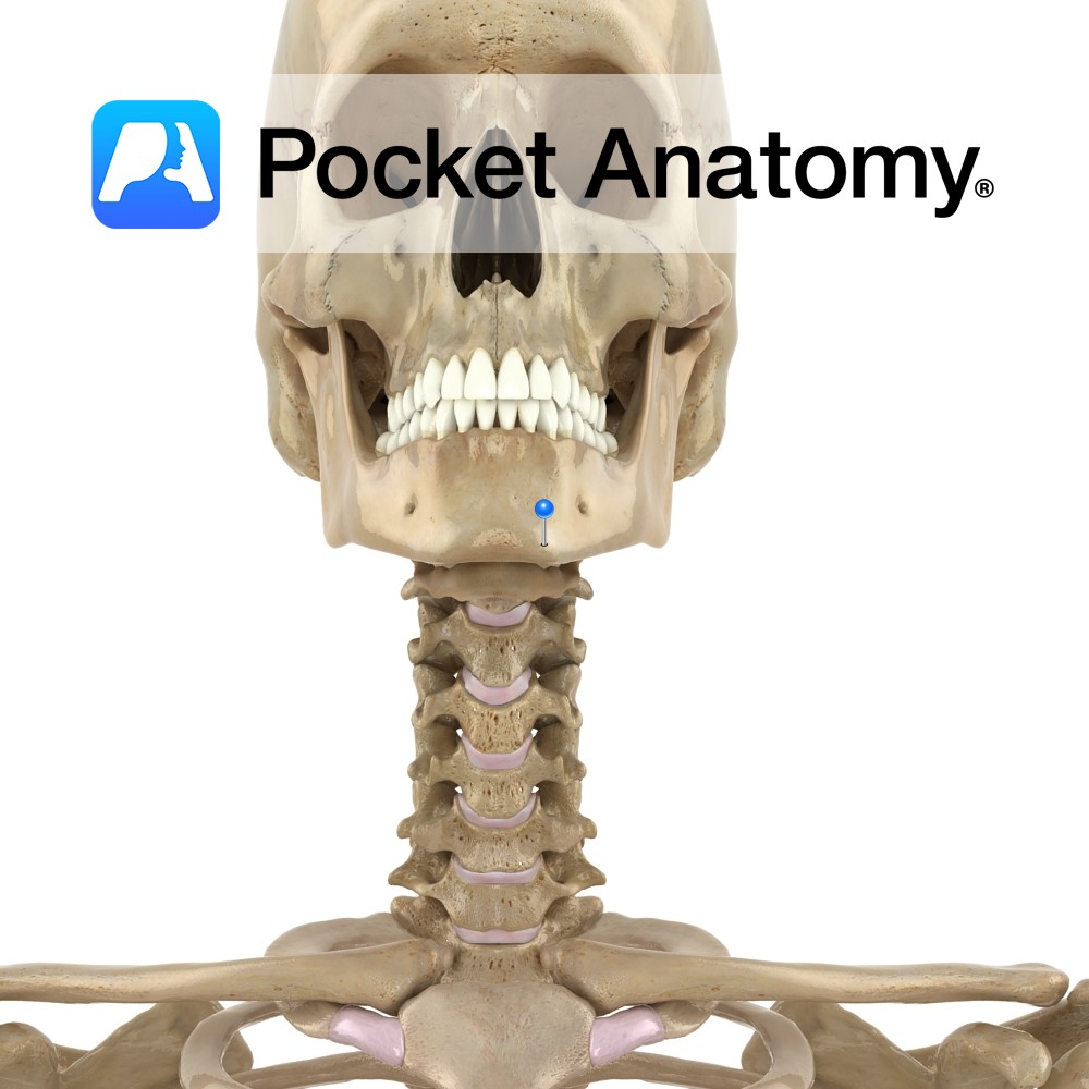
Anatomy The midline symphysis menti marks the fusion of left and right parts of body of mandible. It spreads at its inferior end, forming a forward mental protuberance and on each side, a small mental tubercle. Oblique line ends here. Vignette Chin dimple when there is incomplete fusion of right and left parts of body
- Published in Pocket Anatomy Pins
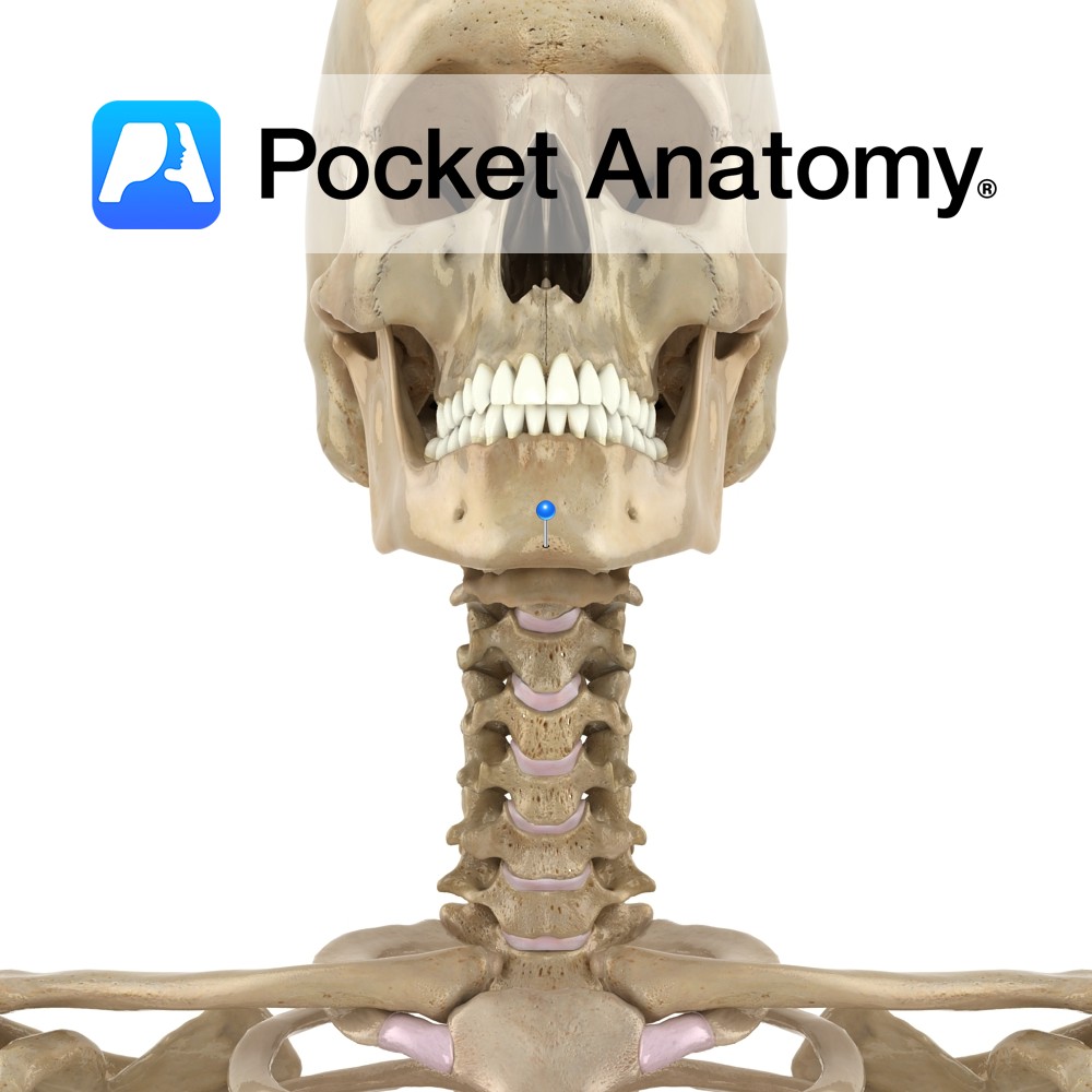
Anatomy The midline symphysis menti marks the fusion of left and right parts of body of mandible. It spreads at its inferior end, forming a forward mental protuberance and on each side, a small mental tubercle. Vignette Chin dimple when incomplete fusion of right and left parts of body of mandible (or of muscle). Inherited
- Published in Pocket Anatomy Pins