PocketAnatomy® is a registered brand name owned by © eMedia Interactive Ltd, 2009-2022.
iPhone, iPad, iPad Pro and Mac are trademarks of Apple Inc., registered in the U.S. and other countries. App Store is a service mark of Apple Inc.
.jpg)
Anatomy Articulates posteriorly with 5th proximal and anteriorly with 5th distal phalanges. Interested in taking our award-winning Pocket Anatomy app for a test drive?
- Published in Pocket Anatomy Pins
.jpg)
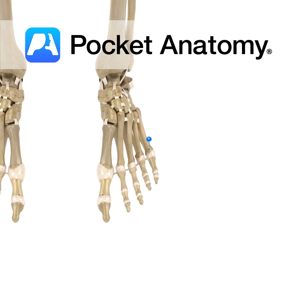
Anatomy Articulates up with 3rd proximal and down with 3rd distal phalanges. Interested in taking our award-winning Pocket Anatomy app for a test drive?
- Published in Pocket Anatomy Pins
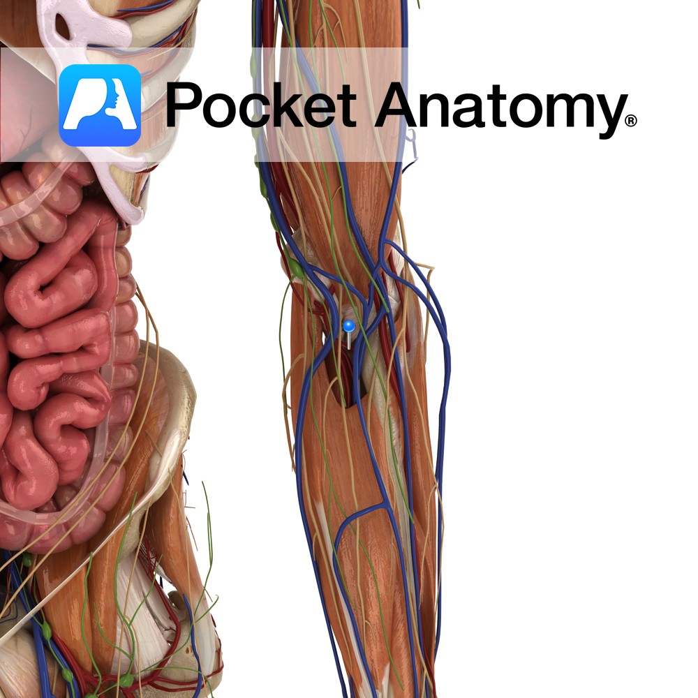
Anatomy Course Branches from the deep brachial artery in the arm, about halfway down the humerus, before the deep brachial artery leaves the radial groove. It then descends to the elbow joint on the posterior aspect of the humerus until it reaches the elbow joint, where it anastomoses with the interosseous recurrent artery. Supply Contributes
- Published in Pocket Anatomy Pins
Motion The metatarsophalangeal joints are ellipsoid type synovial joints formed between the sphere shaped heads of the metatarsal bones and the bases of the proximal phalanges of the toes.The joints allow flexion and extension with restricted abduction, adduction, rotation and circumduction. Stability The fibrous capsule surrounding the joint plays a role in its stability. Ligaments
- Published in Pocket Anatomy Pins
.jpg)
Anatomy On lateral edge foot behind little toe, articulates proximally with cuboid and in (from base) with 4th metatarsal, distally with 5th proximal phalanx. Vignette Metatarsal – tarsal articulations; 1st – medial (1st) cuneiform, 2nd – all 3 cuneiforms, 3rd – lateral (3rd) cuneiform, 4th – lateral cuneiform and cuboid, 5th – cuboid. Interested in
- Published in Pocket Anatomy Pins
.jpg)
Anatomy Articulates proximally with lateral (3rd) cuneiform, out with 4th and in with 2nd metatarsals (from base), distally with 2nd proximal phalanx. Interested in taking our award-winning Pocket Anatomy app for a test drive?
- Published in Pocket Anatomy Pins
.jpg)
Anatomy Thickest shortest metatarsal, on medial edge foot behind/proximal to big toe, articulates proximally with medial (1st) cuneiform, sometimes out (from base) with 2nd metatarsal, distally with 1st proximal phalanx and below (from head) with sesamoid bones. Tibialis anterior, peroneus longus attached. Vignette Metatarsal bones are prismoid, thicker at proximal end (base/ posterior extremity/tarsal end),
- Published in Pocket Anatomy Pins
Motion These are the joints lying between the distal heads of the metacarpals and the proximal phalanges of the digits. They are synovial condylar joints. They have a wide range of motion, including: flexion, extension, abduction and adduction. The flexion component of these is the most extensive, commonly terminated by a grasped object. Stability Articular
- Published in Pocket Anatomy Pins
.jpg)
Anatomy Articulates up with hamate, out/laterally with 4th metacarpal, down with proximal phalanx (5th MCP). Interested in taking our award-winning Pocket Anatomy app for a test drive?
- Published in Pocket Anatomy Pins
.jpg)
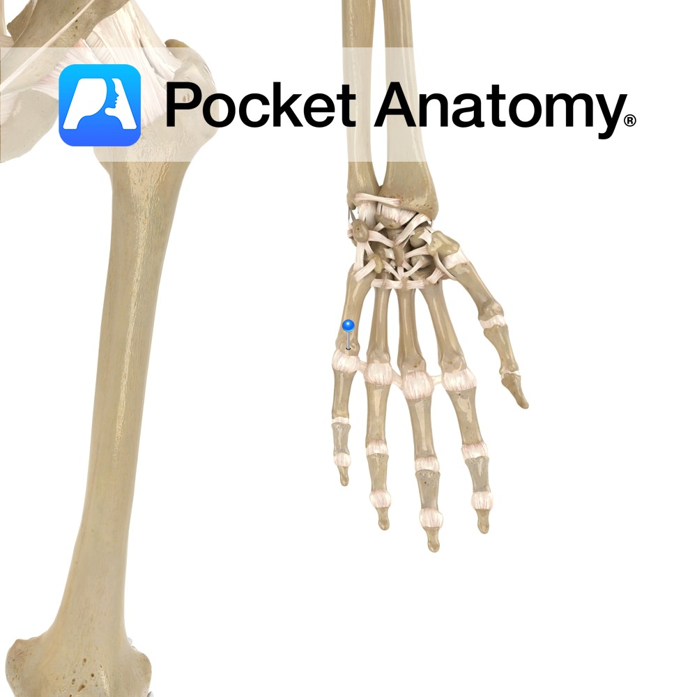
Anatomy Articulates up with capitate, out with index metacarpal, in with ring metacarpal (1st thumb, 2nd index, 3rd middle, 4th ring, 5th little), down with proximal phalanx (3rd MCP). Clinical MCP is a knuckle (4 each hand, as thumb MCP not ordinarily included). Common sites for fractures and other fist injuries. Interested in taking our
- Published in Pocket Anatomy Pins