PocketAnatomy® is a registered brand name owned by © eMedia Interactive Ltd, 2009-2022.
iPhone, iPad, iPad Pro and Mac are trademarks of Apple Inc., registered in the U.S. and other countries. App Store is a service mark of Apple Inc.
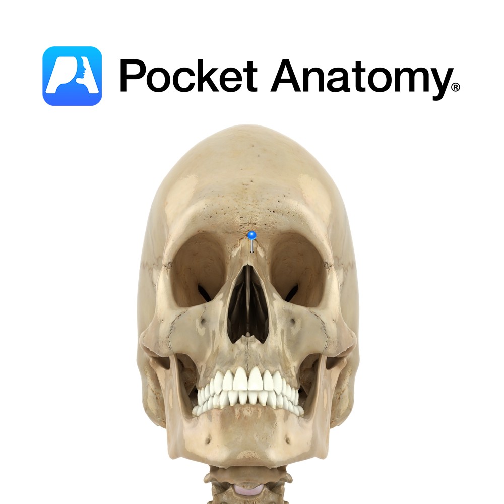
Anatomy Paired, short, oblong. Right and left articulate to form bridge of nose. A crest underneath joint forms part of nasal septum and articulates with spine of frontal, perpendicular plate of ethmoid, and septal cartilage. Each nasal bone articulates with frontal at upper edge, and frontal process of maxilla at lateral edge. Lateral cartilage of
- Published in Pocket Anatomy Pins
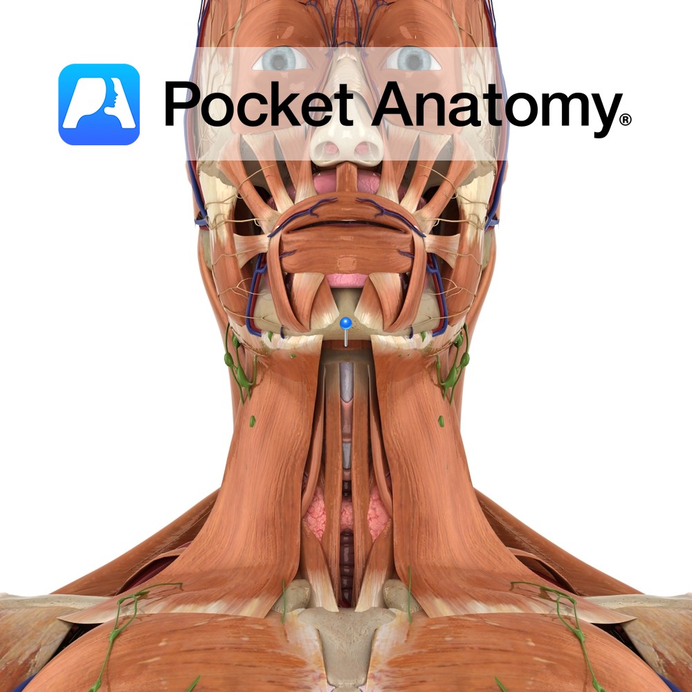
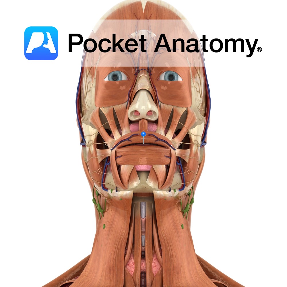
Anatomy Origin: Mylohyoid line on the mandible. Insertion: Body of the hyoid bone and fibres from the muscle on the opposite side. Key Relations: -Is one of the suprahyoid muscles lying in the anterior triangle of the neck. -Forms the floor of the oral cavity with its partner from the opposite side. -Is immediately superior
- Published in Pocket Anatomy Pins
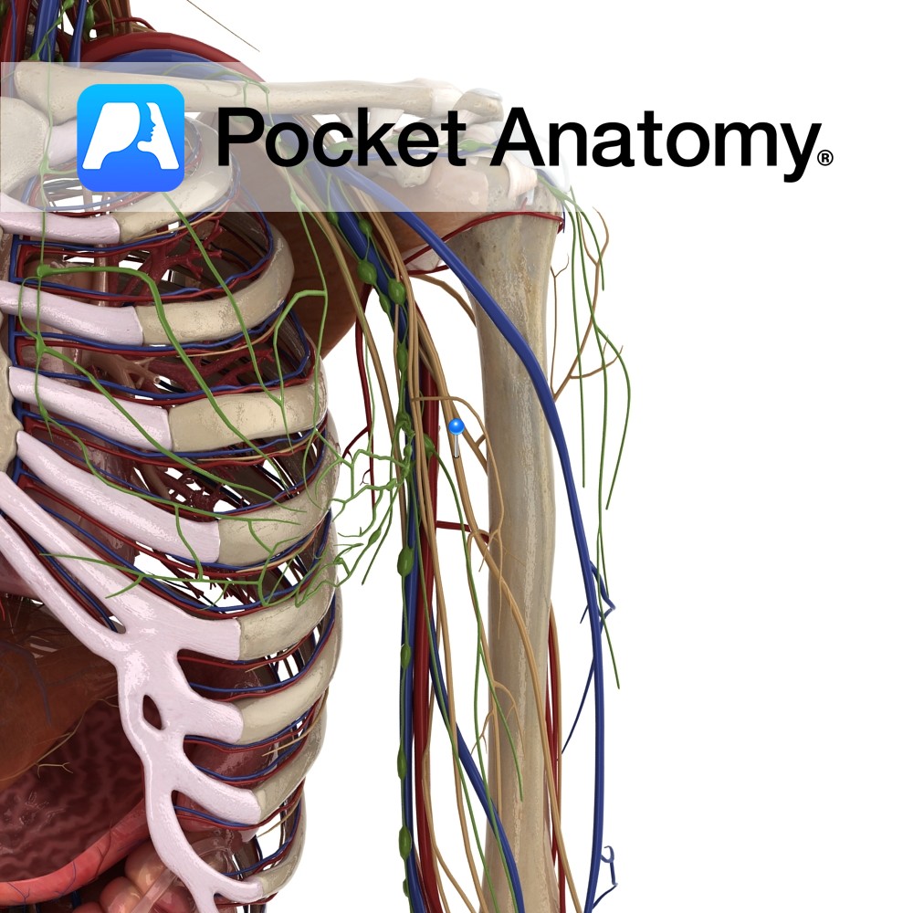
Anatomy Course A terminal branch of the lateral cord of the brachial plexus. It exits the axilla, piercing the coracobrachialis muscle, to pass between the biceps brachii and brachialis muscles in the anterior compartment of the arm. It continues distally towards the elbow, and crosses into the forearm over the lateral epicondyle of the humerus.
- Published in Pocket Anatomy Pins
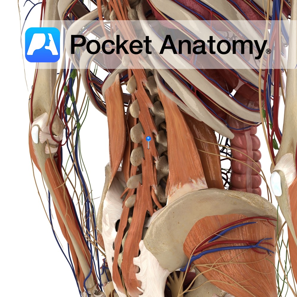
Anatomy One of the intrinsic muscles of the back. Origin: Fasiculi of sacrum, origin of erector spine, posterior superior iliac spine, mamillary processes of lumbar vertebrae, transverse processes of thoracic vertebrae and articular processes of lower four cervical vertebrae. Insertion: Whole length of spinous processes of one of the vertebrae above. Key Relations: Deep to
- Published in Pocket Anatomy Pins
Anatomy Entrance to alimentary tract, a mucomuscular tube from mouth to anus (sight, smell, hunger, anticipation have pre-ingestion roles); oval cavity lower part of head; anterior – lips, lateral – cheeks, inferior – tongue, floor of mouth, superior – hard palate, posterior – oropharynx (starts above at junction hard/soft palates, below behind circumvallate papillae tongue);
- Published in Pocket Anatomy Pins
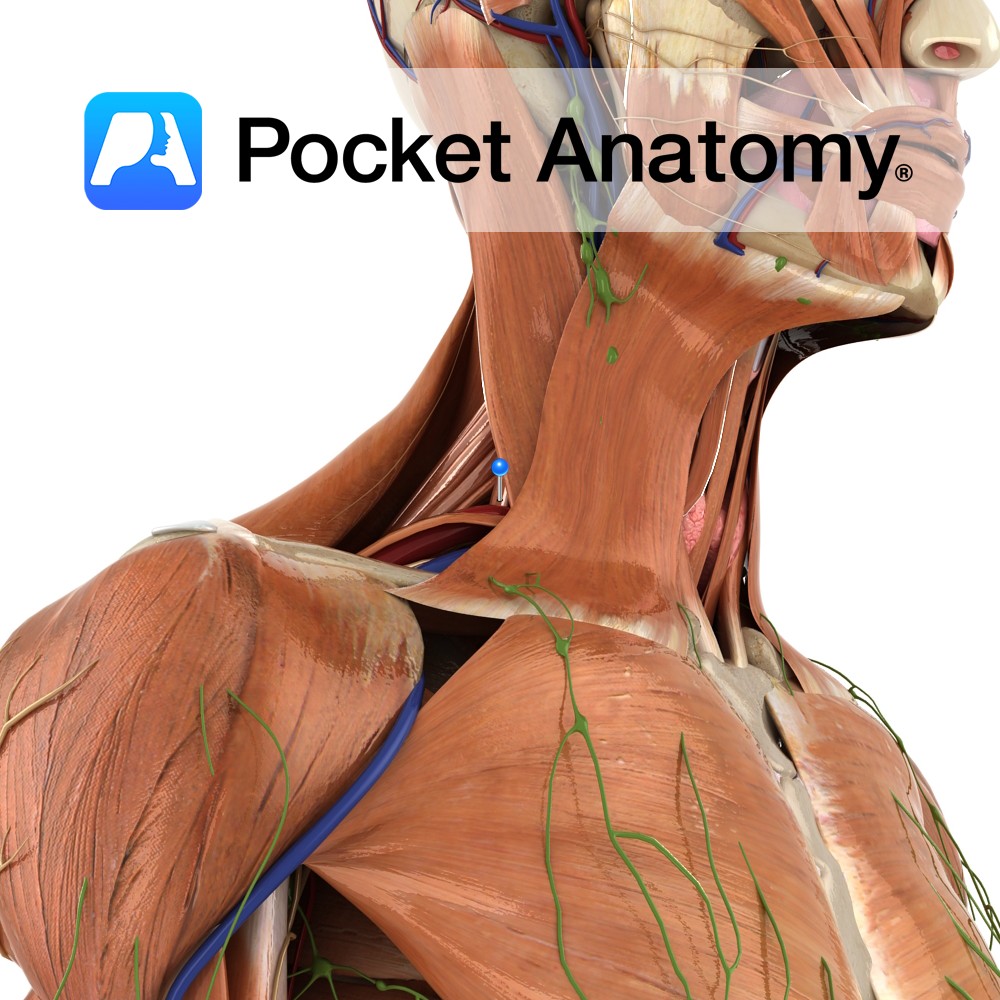
Anatomy Origin: Transverse processes of C2 to C7. Insertion: Upper surface of the 1st rib, posterior to the groove for the subclavian artery. Key Relations: -Largest and longest of the scalenes. -The brachial plexus and the subclavian artery pass between the anterior scalene and the middle scalene. Functions -Elevates the 1st rib. -Rotates the neck
- Published in Pocket Anatomy Pins
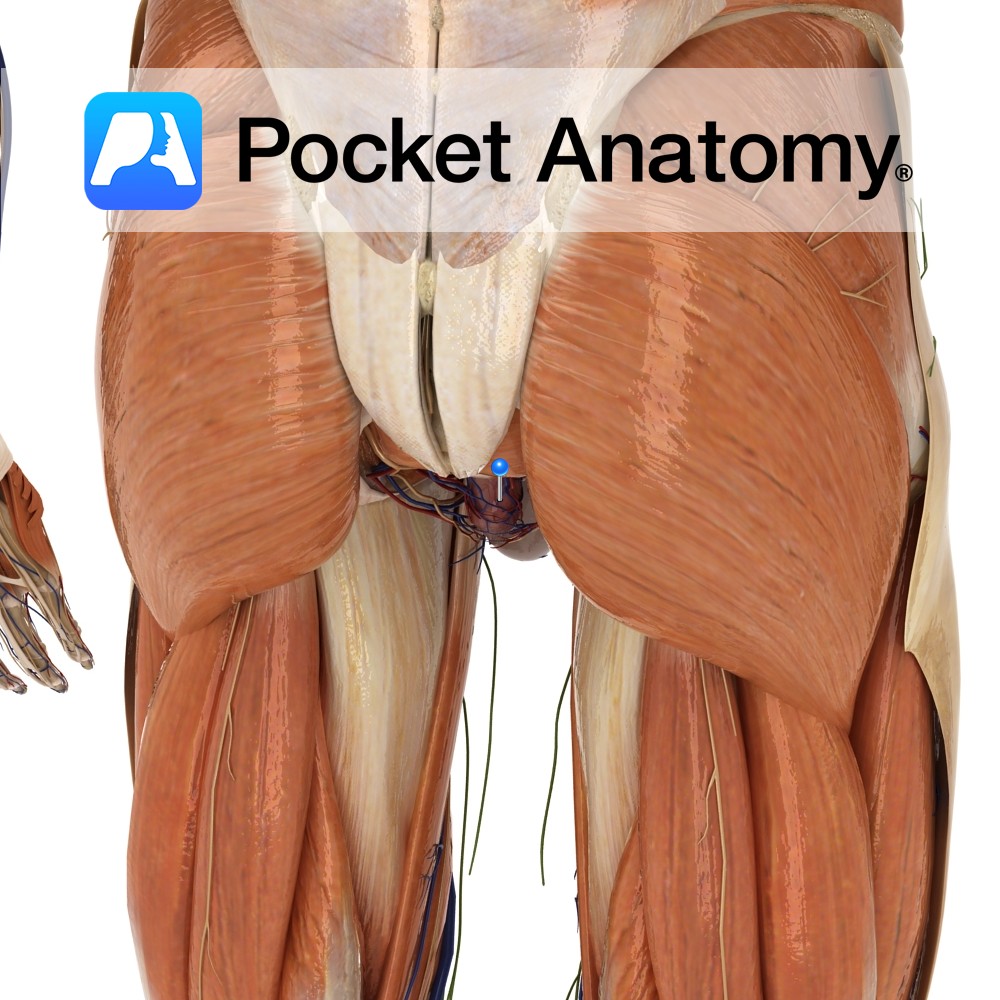
Anatomy Course Originates in the haemorrhoidal plexus of the rectum, courses laterally to drain into the internal iliac vein. Drain Drains the rectum and can receive tributaries from the prostate and occasionally the bladder. Interested in taking our award-winning Pocket Anatomy app for a test drive?
- Published in Pocket Anatomy Pins
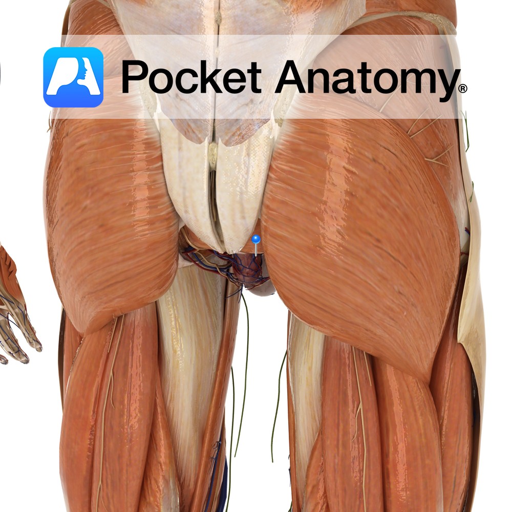
Anatomy Course A branch of the anterior trunk of the internal iliac artery. It branches from the anterior trunk before the anterior trunk passes between the rami anterior rami of two of the sacral nerves. It courses medially along the posterior pelvic wall towards the rectum, where it joins with the superior and inferior rectal
- Published in Pocket Anatomy Pins
.jpg)
Anatomy Articulates up/proximally with proximal phalanx at 5th PIP, and distally with distal phalanx at 5th distal interphalangeal joint (DIP). Interested in taking our award-winning Pocket Anatomy app for a test drive?
- Published in Pocket Anatomy Pins
.jpg)
Anatomy Articulates up/proximally with proximal phalanx at 3rd PIP, and distally with distal phalanx at 3rd distal interphalangeal joint (DIP). Interested in taking our award-winning Pocket Anatomy app for a test drive?
- Published in Pocket Anatomy Pins