PocketAnatomy® is a registered brand name owned by © eMedia Interactive Ltd, 2009-2022.
iPhone, iPad, iPad Pro and Mac are trademarks of Apple Inc., registered in the U.S. and other countries. App Store is a service mark of Apple Inc.
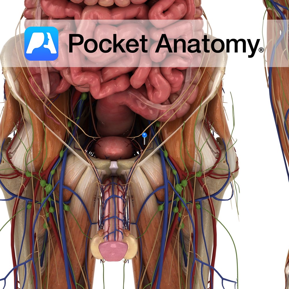
Anatomy Course A branch of the anterior trunk of the internal iliac artery. It branches from the anterior trunk just below the pelvic inlet, and runs anteriorly along the margin of the pelvic inlet. It leaves the pelvic cavity via the obturator canal. As it passes through the obturator canal it splits into two terminal
- Published in Pocket Anatomy Pins
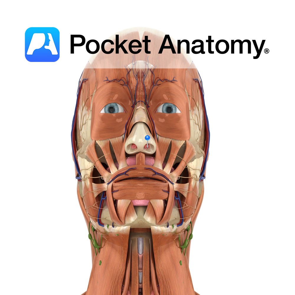
Functions Along with nasal cavity, helps warm and humidify air. Normally first contact body has with cold, external air. It is thought that olfactory ‘perception’ can occur when each nostril is presented with a different smell. Anatomy One of two channels of the nose; where the nasal cavity opens exteriorly. Externally ‘comma’ shaped; usually 7-9mm
- Published in Pocket Anatomy Pins
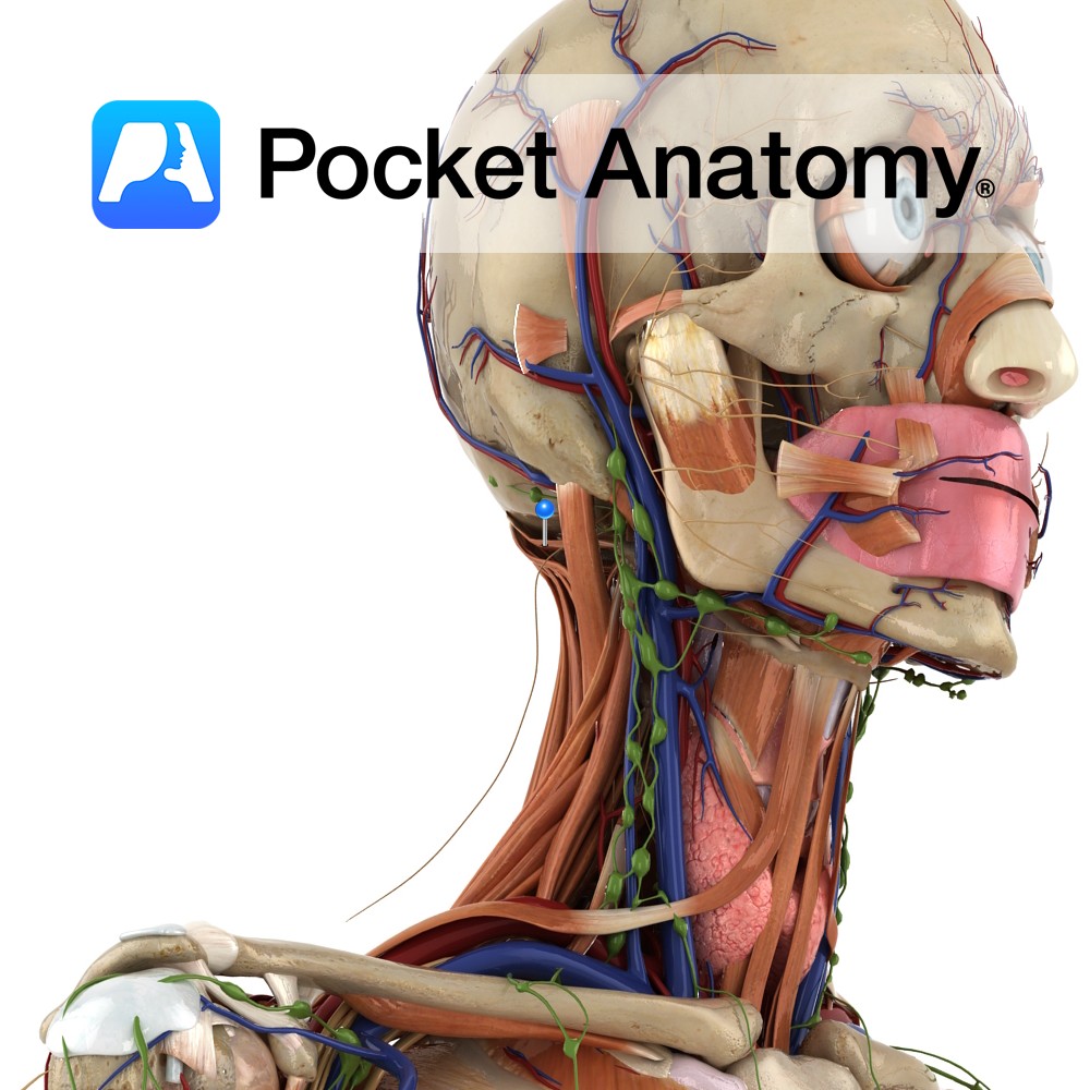
Anatomy Origin: Transverse process of the atlas. Insertion: Lateral half of inferior nuchal line to superior nuchal line. Key Relations: Forms the superior and lateral border of the suboccipital triangle. Functions -Acts bilaterally to extend the head at the atlanto-occipital joint -Acts unilaterally to flex the head to the ipsilateral side. Supply Innervation: Suboccipital nerve
- Published in Pocket Anatomy Pins
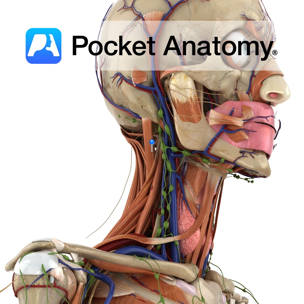
Anatomy Origin: Spinous process of axis. Insertion: Transverse process of the atlas. Key Relations: Forms the inferior and lateral border of the suboccipital triangle. Functions Rotates head to ipsilateral side at atlanto-axial joint. Supply Innervation: Suboccipital nerve (C1). Blood supply: Muscular branches of vertebral artery. Interested in taking our award-winning Pocket Anatomy app for a
- Published in Pocket Anatomy Pins
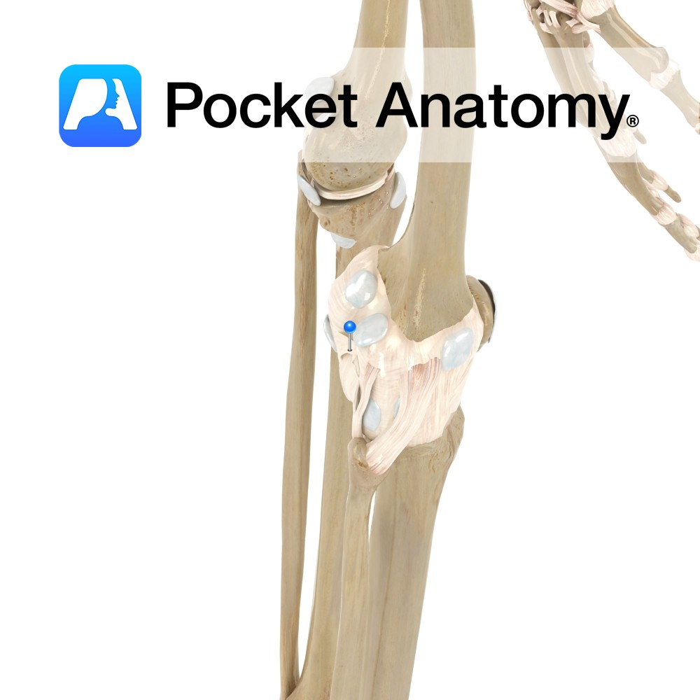
Anatomy A broad, flat ligament forming the floor of the popliteal fossa. This ligament is an extension of the tendon of semimembranosus, attaching from the superior margin of the intercondylar ridge, posterior surface of the femur, and the articular margins of the condyles, down to the posterior margin of the tibia. The ligament reflects across
- Published in Pocket Anatomy Pins
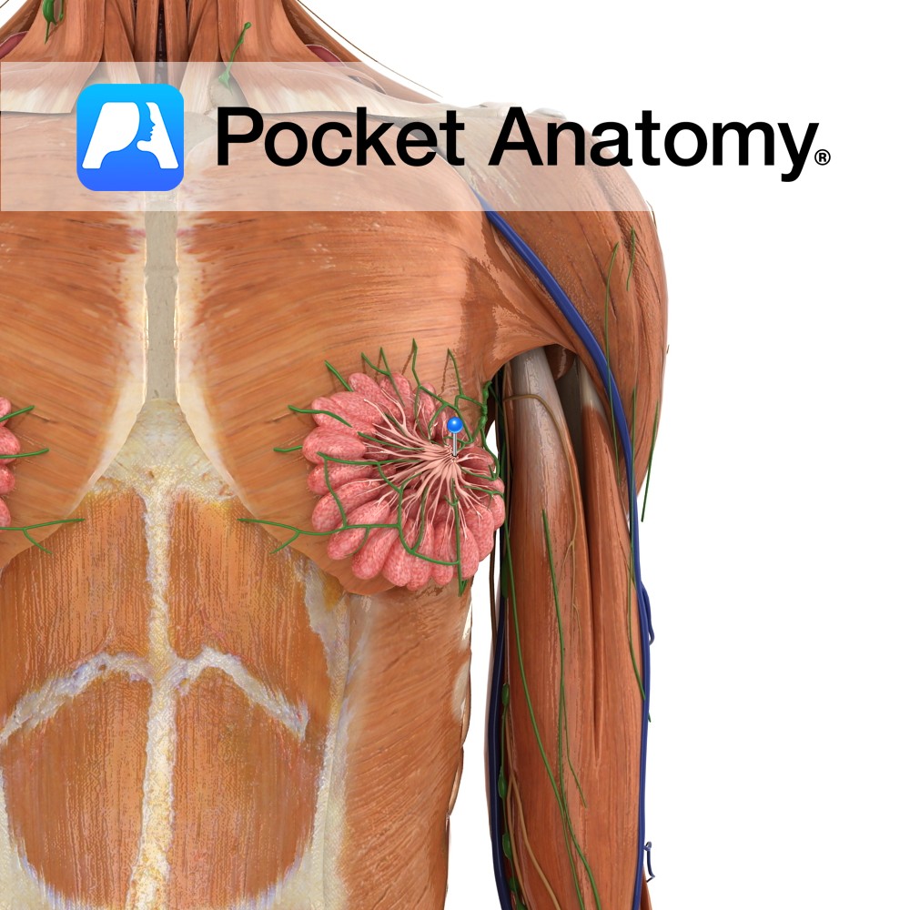
Anatomy (nipple/papilla/teat) Small (about 1cm, size varies, bigger during and subsequent to lactation) skin-covered projection/bump/eminence on front centre of breast (surrounded by sensitive pigmented areola with Montgomery’s tubercles), onto the surface of which project the ends/openings of 5-20 milk ducts from related breast lobules and their acini. Pigmented brown (also areola, more so after pregnancy),
- Published in Pocket Anatomy Pins
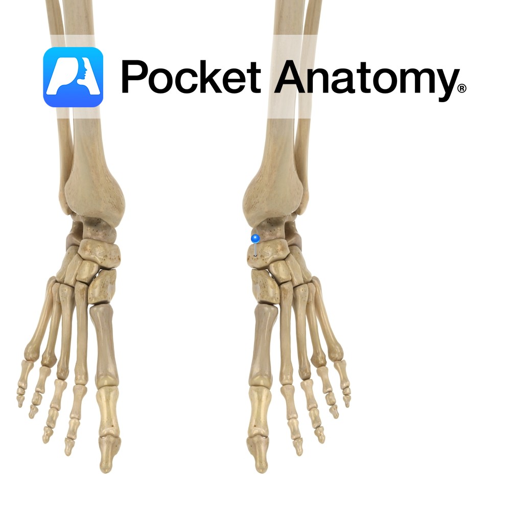
Anatomy Tarsal bone. Articulates up with talus, down/distally with cuneiforms, sometimes out (laterally) with cuboid. Clinical Fracture not evident on plain x-ray, so diagnosis often delayed. Avulsion fracture commonest. Stress fracture best treated early, can result in longterm disabling pain untreated; should be outruled in any young athlete with midfoot pain. Vignette Navicula (Latin); little
- Published in Pocket Anatomy Pins
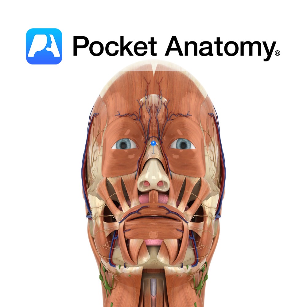
Anatomy Origin: Transverse part: Maxilla just lateral to the nose. Alar part: Maxilla over the lateral incisor. Insertion: Transverse part: Aponeurosis across the dorsum of the nose, with fibres from other side. Alar part: Alar cartilage of the nose. Key Relations: Largest of the muscles in the nasal group. Functions Transverse part: Compresses the nasal
- Published in Pocket Anatomy Pins
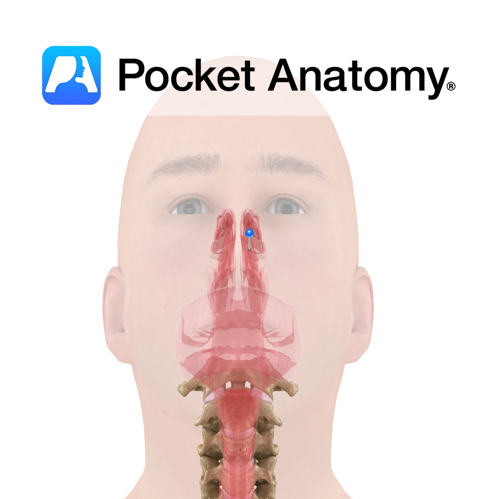
Anatomy Extends from the nostrils to the posterior nasal apertures where the nasopharynx begins. The cavity is pear shaped on the coronal section and divided by a median septum. The borders are as follows: The roof is very narrow (2cm) and forms the floor of the anterior cranial fossa. It is formed by the cribriform
- Published in Pocket Anatomy Pins
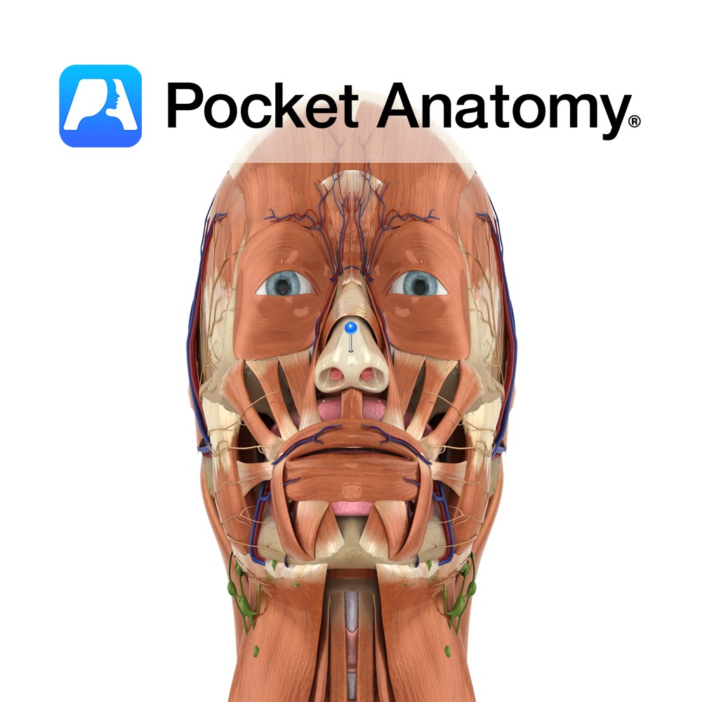
Anatomy Quadrilateral in shape and thicker at its margins than the centre. It attaches to palatine process of the maxilla bone posterosuperiorly, the vomer bone posteroinferiorly, anterosuperiorly it is continuous with the anterior margin of the lateral cartilages and anteroinferiorly connects to the medial crura of the greater alar cartilages by fibrous tissue. Functions Forms
- Published in Pocket Anatomy Pins