PocketAnatomy® is a registered brand name owned by © eMedia Interactive Ltd, 2009-2022.
iPhone, iPad, iPad Pro and Mac are trademarks of Apple Inc., registered in the U.S. and other countries. App Store is a service mark of Apple Inc.
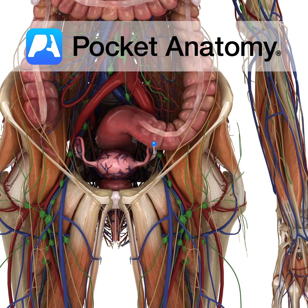
Anatomy Female gonad (organ which secretes gametes – male/female secretes sperm/eggs). Paired, 4X2X1cm, 2-3.5gm (smaller with age from puberty), almond-shaped (long axis vertical), grey-pink, surface smooth (or puckered after repeated ovulation), sits at side of uterus, just below uterine/fallopian tube, in pelvic cavity, at or below level of umbilicus, in shallow ovarian fossa in lateral
- Published in Pocket Anatomy Pins
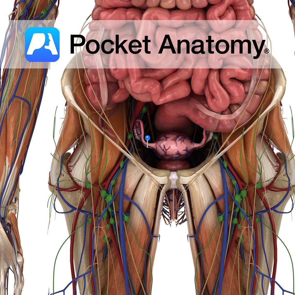
Anatomy Fibromuscular ligament connecting the lower/uterine extremity of the ovary to the side of the uterus, near the fundus and the uterotubal junction. Clinical Not to be confused with the suspensory ligament of ovary, a part of the upper edge of the broad ligament which connects the other, upper, tubal extremity of the ovary to
- Published in Pocket Anatomy Pins
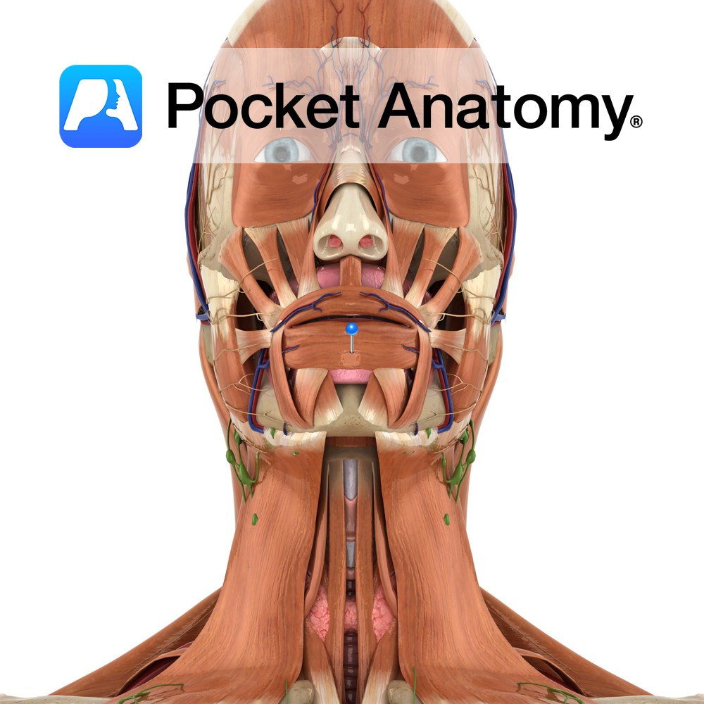
Anatomy Origin: Mandible below and maxilla above, some fibres originate from other muscles of the face (buccinator, depressor anguli oris, zygomaticus major and minor, quadratus labii inferioris and superioris) Insertion: The skin of the lips and the mucous membrane beneath the lips. Key Relations: Fibres completely encircle the mouth. Functions -Acts in the manner of
- Published in Pocket Anatomy Pins
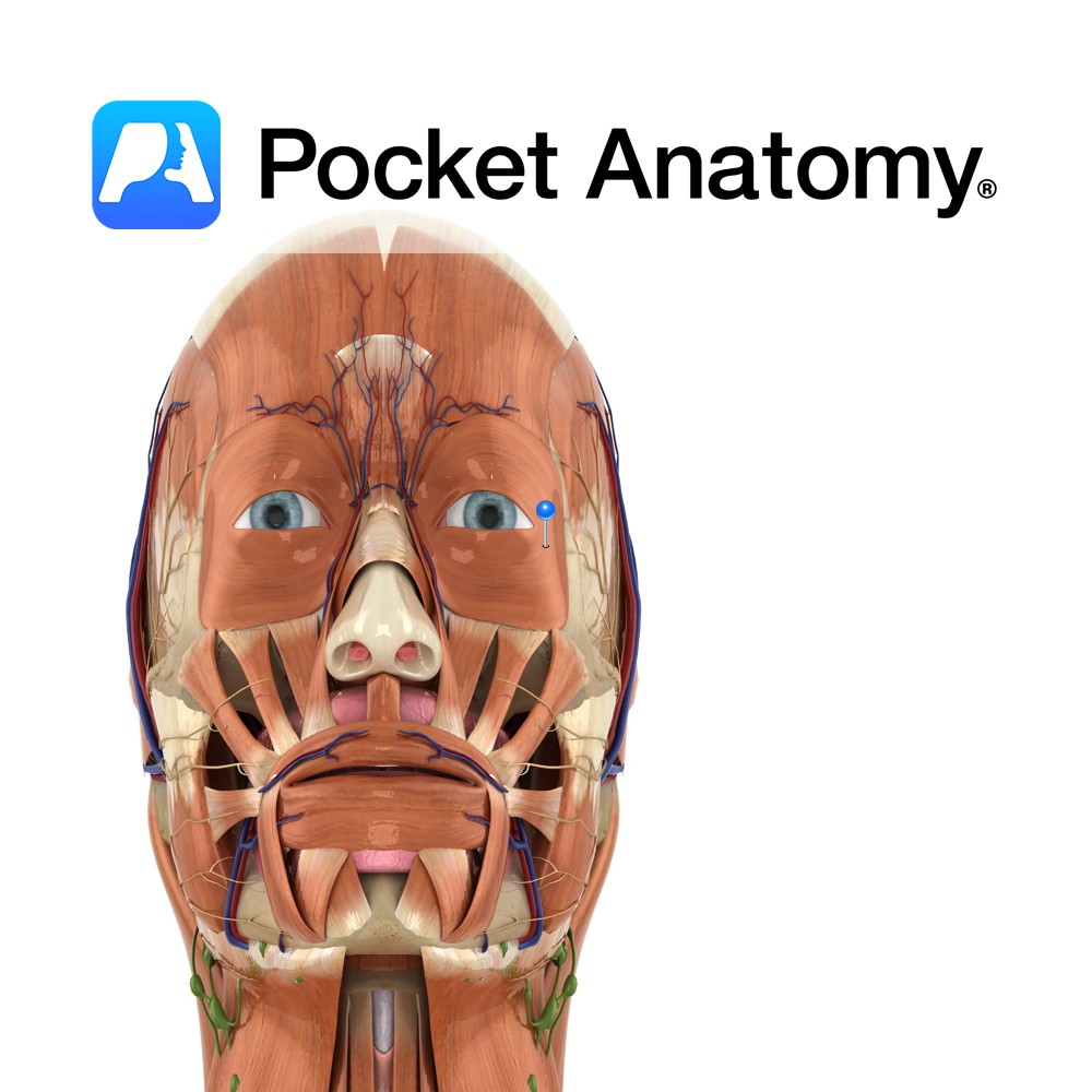
Anatomy Origin: Palebral part: Medial palpebral ligament. Orbital part: Nasal portion of frontal bone, frontal process of maxilla, medial palebral ligament. Insertion: Palebral part: Lateral palebral raphe at outer corner of eye. Orbital part: Fibres converge and form uninterrupted elipse around orbit of the eye. Key Relations: A third part of the muscle exists known
- Published in Pocket Anatomy Pins
.jpg)
Anatomy Origin: Optic nerve fibers are the axons of the cells in the ganglionic layer of the retina. Structure: The three sheaths surrounding the nerve are continuous with the meninges. The outermost sheath originates from the dura mater and merges with the sclera of the eyeball, the middle sheath is continuous with the arachnoid mater
- Published in Pocket Anatomy Pins
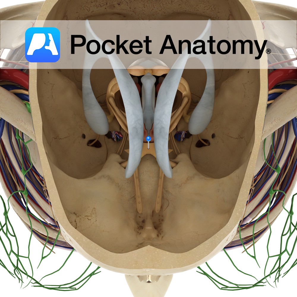
Anatomy The two optic nerves unite at the optic chiasm which is located at the base of the brain in the interpeduncular cistern. It is situated between the two internal carotid arteries, and the tuber cinereum of the hypothalamus lies posterior to the chiasm. Blood Supply: Supplied by the branches of many vessels, particularly the
- Published in Pocket Anatomy Pins
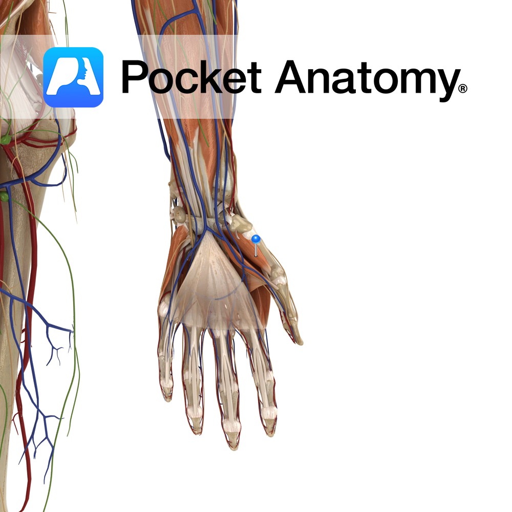
Anatomy Origin: Tubercle of trapezium and flexor retinaculum. Insertion: Radial border and adjacent palmar surface of the first metacarpal (thumb). Key Relations: Is one of the muscles of the thenar eminence of the hand. Functions Flexion, abduction, and medial rotation (i.e. the action of opposition) of the metacarpal of the thumb at the carpopmetacarpal and
- Published in Pocket Anatomy Pins
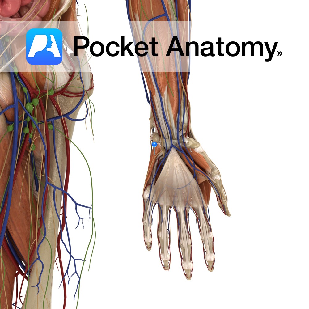
Anatomy Origin: Hook of the hamate and the flexor retinaculum. Insertion: Ulnar aspect of the fifth metacarpal. Key Relations: Is one of the muscles of the hypothenar eminence of the hand. Functions Flexion and lateral rotation (i.e.opposition) of the fifth metacarpal at the carpometacarpal and metacarpophalangeal joint. Supply Nerve Supply: Deep branch of the ulnar
- Published in Pocket Anatomy Pins
.jpg)
Anatomy Course: The superior and inferior ophthalmic veins begin on their respective sides of the eye. They drain backwards towards the cranial cavity, merging to form a single vessel that empties into the cavernous sinus. Supply Drains the orbit of the eye and its structures. Interested in taking our award-winning Pocket Anatomy app for a
- Published in Pocket Anatomy Pins