PocketAnatomy® is a registered brand name owned by © eMedia Interactive Ltd, 2009-2022.
iPhone, iPad, iPad Pro and Mac are trademarks of Apple Inc., registered in the U.S. and other countries. App Store is a service mark of Apple Inc.
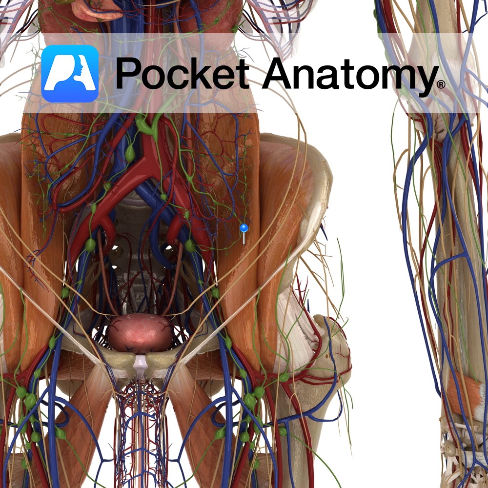
Anatomy Origin: Sides of vertebra T12 to L5 and transverse processes of L1 to L5. Insertion: Lesser trochanter of the femur. Key Relations: -The ureter passes along the medial aspect of the psoas major muscle as it enters the pelvis. -Forms the floor of the femoral triangle. -The femoral nerve (L2 to L4) travels through
- Published in Pocket Anatomy Pins
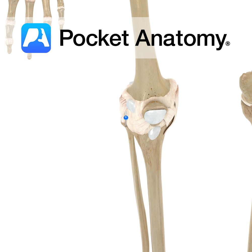
Motion The proximal tibiofibular joint is a synovial plane joint. The lateral condyle of the tibia articulates with the articular facet of the head of the fibula. It allows very little movement. Slight movement occurs to accommodate the movement of the medial malleolus and fibula during plantar or dorsiflexion of the foot. Stability The fibrous
- Published in Pocket Anatomy Pins
.jpg)
Anatomy Articulates up with metacarpal at 5th MCP, down with middle phalanx at proximal interphalangeal joint (PIP). Clinical Rheumatoid arthritis commoner MCPs and PIPs, osteoarthritis commoner DIPs. Interested in taking our award-winning Pocket Anatomy app for a test drive?
- Published in Pocket Anatomy Pins
.jpg)
Anatomy Articulates up with metacarpal at 3rd MCP, down with middle phalanx at proximal interphalangeal joint (PIP). Interested in taking our award-winning Pocket Anatomy app for a test drive?
- Published in Pocket Anatomy Pins
.jpg)
Anatomy At near end is 1st MCP. Thumb has only 2 phalanges, joint between is intermediate interphalangeal. Interested in taking our award-winning Pocket Anatomy app for a test drive?
- Published in Pocket Anatomy Pins
.jpg)
Anatomy Articulates posteriorly with 5th metatarsal, anteriorly with 5th middle phalanx. Interested in taking our award-winning Pocket Anatomy app for a test drive?
- Published in Pocket Anatomy Pins
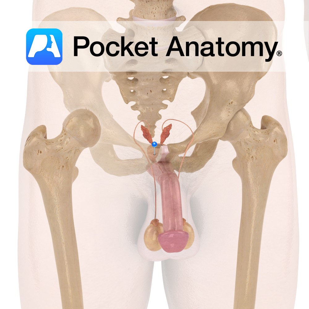
Functions Secretes alkaline fluid which constitutes ~30% of semen (with spermatozoa and seminal vesicle fluid) and protects sperm against acidic vaginal environment. This fluid is produced constantly, with the excess being expelled in urine. Contains smooth muscles which help project sperm during ejaculation. These muscles also help in involuntary control of urine. Anatomy Complex exocrine
- Published in Pocket Anatomy Pins
.jpg)
Anatomy Articulates proximally with 3rd metatarsal, distally with 3rd middle phalanx. Interested in taking our award-winning Pocket Anatomy app for a test drive?
- Published in Pocket Anatomy Pins
.jpg)
Anatomy Articulates up with 1st metatarsal, down with 1st distal phalanx (big toe has only 2 phalanges). Interested in taking our award-winning Pocket Anatomy app for a test drive?
- Published in Pocket Anatomy Pins

Anatomy Course Begin at the metacarpals and drain blood into the venous palmar arch of the hand. Drain Drain into the palmar venous plexus. Interested in taking our award-winning Pocket Anatomy app for a test drive?
- Published in Pocket Anatomy Pins