PocketAnatomy® is a registered brand name owned by © eMedia Interactive Ltd, 2009-2022.
iPhone, iPad, iPad Pro and Mac are trademarks of Apple Inc., registered in the U.S. and other countries. App Store is a service mark of Apple Inc.
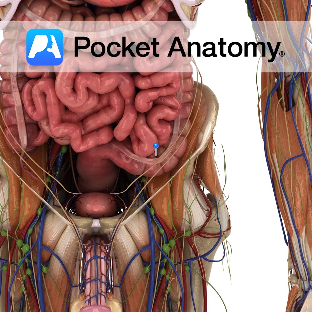
Anatomy Loop of colon continuous above with descending colon, ordinarily within pelvis (mobile, can be displaced), starting at pelvic brim, passing across in front of sacrum from left to right, then back left to midline at level S3 (3rd fused sacral vertebra), then down to be continuous with rectum, surrounded by peritoneum with a mesocolon,
- Published in Pocket Anatomy Pins
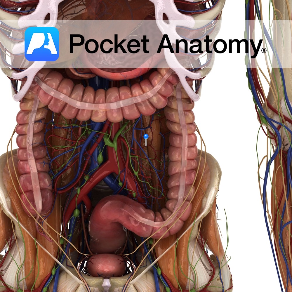
Anatomy Course There can be two to four sigmoid arteries. These are branches from the inferior mesenteric artery that descend towards the sigmoid colon in the lower left quadrant of the abdomen. They travel within the sigmoid mesocolon and anastomose with branches from the left colic artery and the superior rectal artery. Supply Supply sigmoid
- Published in Pocket Anatomy Pins
.jpg)
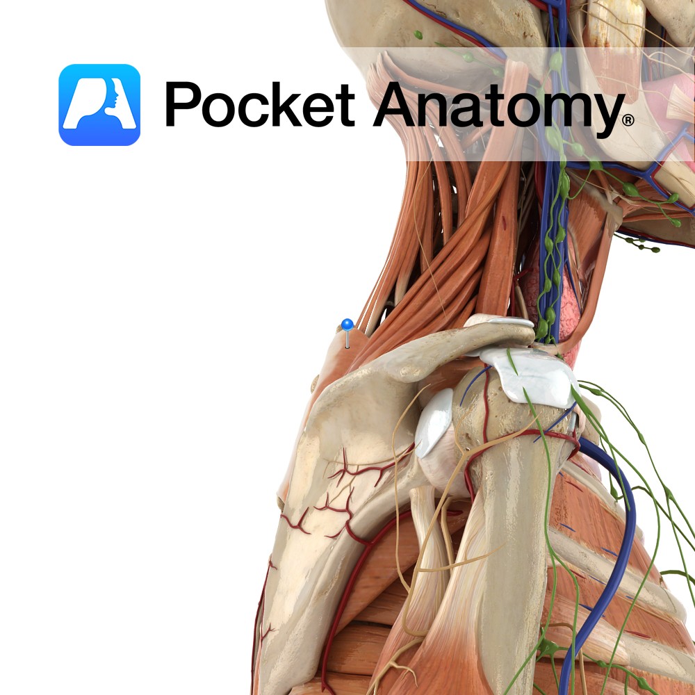
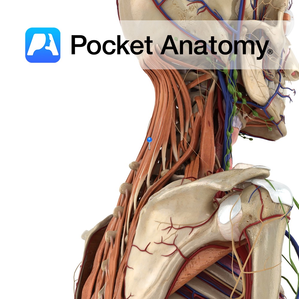
Motion The glenohumeral joint is a multiaxial synovial ball and socket joint and involves the articulation between the glenoid fossa of the scapula and the head of the humerus. The glenohumeral joint is the most mobile joint in the body. It is capable of flexion, extension, abduction, adduction, medial rotation, lateral rotation, and circumduction. Circumduction
- Published in Pocket Anatomy Pins
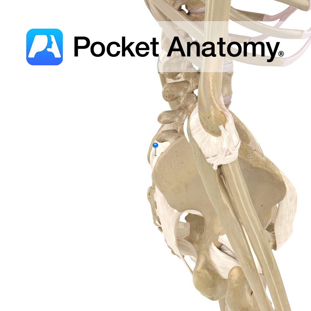
Anatomy Horizontal in direction, attaching from the first and second transverse tubercles on the posterior surface of the sacrum to the tuberosity of the ilium. Functions Supplies static stability to the sacroiliac joint. Interested in taking our award-winning Pocket Anatomy app for a test drive?
- Published in Pocket Anatomy Pins
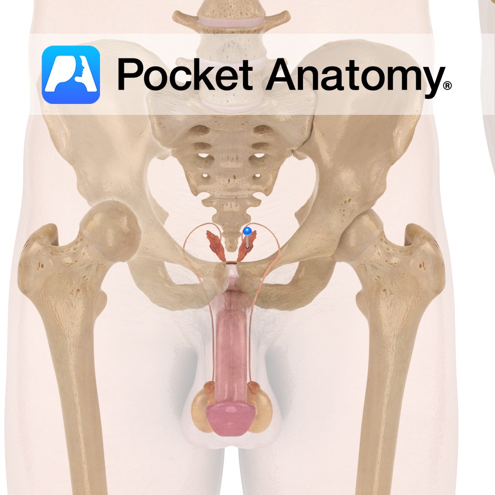
Functions Creates a significant portion (50-70%) of seminal fluid, though not all is released in the primary ejaculation (which consists mainly of prostatic fluid). The seminal vesicle empties into the vas deferens. (Note that the seminal vesicle forms in utero from a pouch of the vas deferens.). Anatomy Pair of tubular glands attached close to
- Published in Pocket Anatomy Pins
Anatomy Origin: Ligamentum nuchae and spinous processes of C7 to T3 and supraspinous ligaments. Insertion: Upper border of 2nd to the 5th ribs just lateral to their angles. Key Relations: -Serratus posterior superior lies superior to the thoracolumbar fascia. -Sometimes referred to as part of the respiratory group. Functions -Elevates 2nd to the 5th ribs,
- Published in Pocket Anatomy Pins
Anatomy Origin: Spinous processes of T11 to L3 and supraspinous ligaments. Insertion: Lower border of the 9th to the 12th ribs just lateral to their angles. Key Relations: -Serratus posterior inferior shares it’s aponeurosis of origin with latissimus dorsi. -Sometimes referred to as part of the respiratory group. Functions -Depresses 9th to the 12th ribs,
- Published in Pocket Anatomy Pins
Anatomy Origin: External surface of lateral surface of upper eight ribs. Insertion: Medial border of scapula. Key Relations: The long thoracic nerve travels inferiorly on the surface of the muscle. Functions -Protracts the scapula laterally rotates the scapula e.g. as in the forward motion of throwing a punch. -Stabilises and holds the medial border of
- Published in Pocket Anatomy Pins
Anatomy Origin: Superomedial area of ischial tuberosity (with biceps femoris). Insertion: Superior part of medial tibia. Key Relations: -One of the three muscles of the posterior compartment of the thigh. -Along with semimembranosus forms the superomedial boundary of the popliteal fossa. -At its insertion to the tibia, the tendon of semitendonosus is located just posterior
- Published in Pocket Anatomy Pins
Anatomy One of the intrinsic muscles of the back. Consists of semispinalis capitis, cervicus and thoracis. Semispinalis capitis: Origin: Articular processes of C4 to C6 and transverse processes of C7 to T7. Insertion: Occipital bone on the area lying between the superior and inferior nuchal lines. Semispinalis cervicis: Origin: Transverse processes of T1 to T5/6.
- Published in Pocket Anatomy Pins