Anatomy
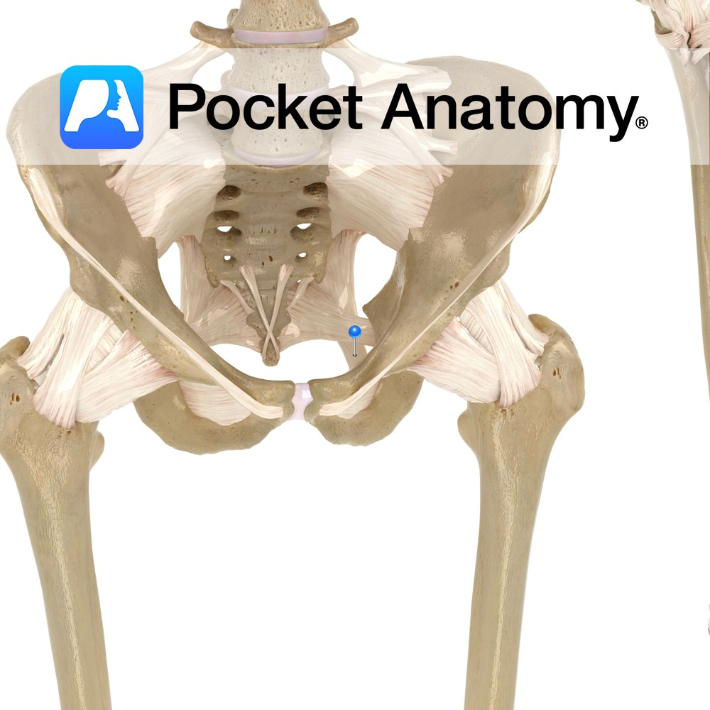
Origin:
Anterior superior iliac spine of hip bone and superior part of notch below it.
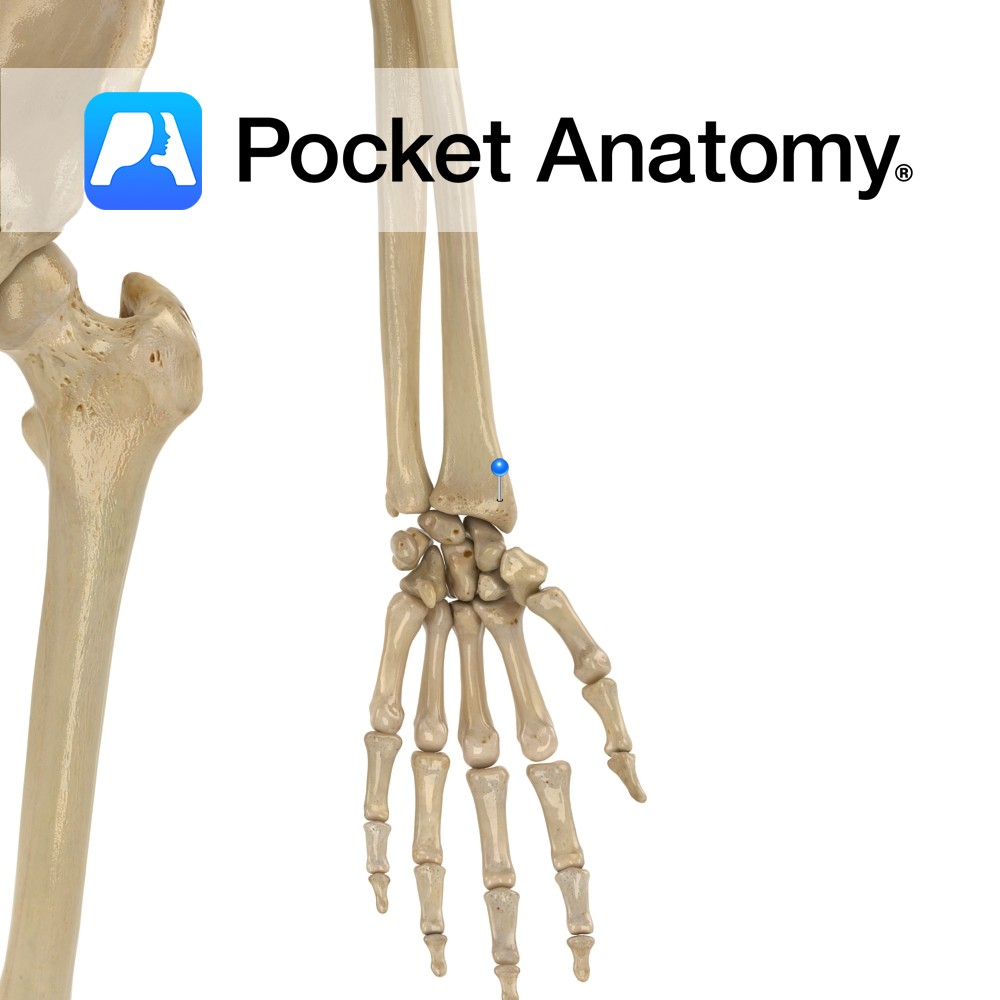
Insertion:
Attaches by aponeurosis to medial surface of the tibia.
Key Relations:
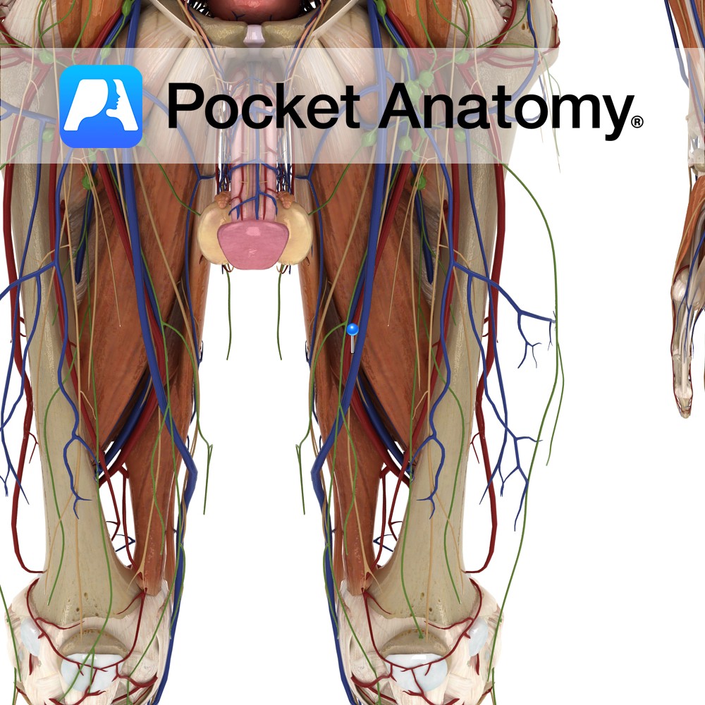
-One of the five muscles of the anterior compartment of the thigh.
-In the upper portion of the thigh sartorius forms the lateral margin of the femoral triangle.
-In the middle portion of the thigh sartorius forms the anterior wall of the adductor canal.
-At its insertion to the tibia, the tendon of sartorius is located just anterior to the tendons of the gracillis and semitendinosus muscles. This 3 pronged arrangement is known as pes anserinus (the latin for goosefoot).
Functions
-Weakly flexes the thigh at the hip joint.
-Abducts and lateral rotates the thigh at the hip joint.
-Flexes the leg at the knee joint.
-Medially rotates leg when knee is flexed.
*Checking the sole of your shoe to see if you stood on something demonstrates all of actions of sartorius.
Supply
Nerve Supply:
Femoral nerve (L2, L3).
Blood Supply:
Femoral artery.
Clinical
The sartorius muscle may be strained particularly by overextending the hip.
Surgeons may use the sartorius muscle when treating complex femoral artery wounds. Placement of healthy soft tissue with intact blood supply into the wound location is known to reduce the incidence of infection and improve wound healing.
Interested in taking our award-winning Pocket Anatomy app for a test drive?
![]()